Syeda Khadija1*, Syed Amir Gilani2, Mohammad1, Sabir Butt1, Raham Bacha1, Syed Zain ul Hassan Gilani1
1Department of Radiological Sciences and Medical Imaging Technology, University of Lahore, Lahore, Pakistan
2Faculty of Allied Health Sciences, University of Lahore, Lahore, Pakistan
*Corresponding Author: Syeda Khadija, Department of Radiological Sciences and Medical Imaging Technology, University of Lahore, Lahore, Pakistan
Received: 13 December 2019; Accepted: 26 December 2019; Published: 02 January 2020
Objective: To identify the usefulness of Doppler indices in the third trimester for intra-uterine growth- restricted pregnancies.
Materials and Methods: It was an observational case-control study of singletons with intra-uterine growth restriction (fetal weight <10th percentile). Intra-uterine growth restricted fetuses (cases) and normal (controls) were examined for the umbilical artery. Time intervals between progressive Doppler abnormalities and configurations of worsening were related to umbilical artery Doppler status and gestational age. This study was conducted from August 2015 to January 2016 at Gilani Ultrasound Center Lahore Pakistan. A total of 60 pregnant females were studied with normal and abnormal umbilical artery Doppler ultrasound.
Results: Doppler indices measurements of umbilical artery in intra-uterine growth restriction fetuses in the third trimester showed higher values as compared to normal fetuses. 30 females had normal umbilical artery Doppler indices and waveforms and 30 females showed (either thinning, absent or reversed Diastolic flow Doppler waveform) with higher indices.
Conclusion: Fetal umbilical artery Doppler ultrasound is an effective tool in the detection of early intra-uterine growth restriction fetuses.
Intrauterine growth restriction, Umbilical artery, Doppler indices (Pulsatility index, resistive index, S/D ratio)
1. Introduction
Intrauterine growth restriction is a sonographic estimated fetal weight which is <10th percentile for gestational age [1]. Bloodstream in the umbilical cord is typically high flow and low resistance with the increased end-diastolic flow as gestational age advances [2]. Color Doppler evaluation plays an imperative role in pregnancy and widely acknowledged functional methods of evaluating fetal wellbeing. Flow velocity waveforms provide essential information from the early stages of pregnancy to term. Umbilical arterial Doppler waveforms reflect the status of the placental course, and an increase in end-diastolic stream that is seen with advancing gestation is a direct result of an increase in the number of tertiary stem villi that takes place with placental maturation [3]. Diseases that obliterate small muscular arteries in placental tertiary stem villi result in a progressive decrease in end-diastolic flow in the umbilical arterial Doppler waveforms until absent, and then reverse flow during diastole is noted [4]. Reversed diastolic flow in the umbilical arterial circulation represents an advanced stage of placental compromise, and is associated with more than 70% of placental arterial obliteration [5]. The absent or reversed end diastolic flow in the umbilical artery is commonly escorting with severe intrauterine growth restriction and oligohydramnios [6]. The cerebral circulation is normally a pronounced impedance circulation with the continuous forward flow throughout the cardiac cycle [7]. The middle cerebral artery is the most accessible, cerebral vessel to ultrasound imaging in the fetus, and it brings more than 80% of cerebral blood flow [8]. In fetal hypoxemia, central redistribution of blood flow occurs, resulting in increased blood flow to the brain, heart, and adrenals, and a lessening inflow to the peripheral and placental circulations. This blood flow redistribution is known as the brain-sparing reflex, and plays an important role in fetal adaptation to deprivation [9]. Arterial Doppler abnormalities, at the level of the umbilical and middle cerebral arteries (brain-sparing reflex), confirm the presence of hypoxemia in intra-uterine growth restriction fetuses, it represents early cautioning signs [10]. The aim of this study was to Doppler velocimetry has enhanced our diagnosis that the occurrence of such abnormal late stage changes of vascular adaptation by the (intra-uterine growth restriction) fetuses appears to be the best predictor of perinatal mortality, and morbidity independent of gestational age and weight.
2. Materials and Methods
Study was conducted after approval from institutional review board. 60 cases of normal and suspected intra-uterine growth restriction visited us during study interval were included. Toshiba Nemio-30 with convex transducer having frequency 3. 5-5 MHZ used for scanning the patients. Before the examination procedure was explained to each patient and informed consent was obtained. The abdomen-o-pelvic area was uncovered, transmitting gel was applied. The transducer was placed over the Para uterine region and free-floating umbilical cord was found on real-time gray scale ultrasound then applied color Doppler and Pulsed Doppler. The superlative setting was pragmatic on pulsed Doppler to avoid aliasing. The sample volume was selected at mid cord level and the waveform was gained at least 2-3 times per patient. Doppler indices were deliberate. Pregnant ladies with singleton pregnancies in the third trimester of pregnancies with certain last menstrual dates were recruited. All fetuses with differences of less than 3 standard deviations in their biometric parameters were included. The indices (PI, RI and SD ratio) were recorded and diastolic flow on the spectral waveform as decreased, absent or reversal of flow. Collected information was entered into the statistical package of the social sciences for analyzed.
3. Results
In this study total 60 pregnant females were recruited and Doppler indices measurements were obtained.
|
N=60 |
Normal/IUGR |
Mean |
Std. Deviation |
|
Age in years |
Normal |
26. 07 |
4. 051 |
|
IUGR |
26. 13 |
4. 637 |
|
|
Gestational age in weeks |
Normal |
33. 4333 |
2. 37753 |
|
IUGR |
33. 69 |
3. 25866 |
|
|
Pulsatility Index (PI) |
Normal |
0. 897 |
0. 2062 |
|
IUGR |
1. 824 |
1. 47869 |
|
|
Resistive Index (RI) |
Normal |
0. 5827 |
0. 09176 |
|
IUGR |
0. 8517 |
0. 22485 |
|
|
Systolic to Diastolic ratio (S/D) |
Normal |
2. 426 |
0. 41359 |
|
IUGR |
3. 8013 |
0. 66226 |
Table 1: Mean values of Doppler indices of normal n IUGR.
|
IUGR-Normal |
Total |
||||
|
absent diastole flow |
decrease diastole flow |
Normal |
|||
|
G-Age (Binned) |
<= 30. 00 |
2 |
2 |
2 |
6 |
|
30. 01 - 33. 00 |
4 |
5 |
11 |
20 |
|
|
33. 01 - 36. 00 |
2 |
7 |
11 |
20 |
|
|
36. 01 - 39. 00 |
4 |
3 |
6 |
13 |
|
|
39. 01+ |
0 |
1 |
0 |
1 |
|
|
Total |
12 |
18 |
30 |
60 |
|
Table 2: Normal and IUGR cross tabulation with G. A.
|
IUGR- Normal |
Total |
||||
|
absent diastole flow |
decrease diastole flow |
Normal |
|||
|
PI (Binned) |
<= 1. 00 |
0 |
0 |
23 |
23 |
|
1. 01 - 2. 00 |
12 |
15 |
7 |
34 |
|
|
4. 01 - 5. 00 |
0 |
1 |
0 |
1 |
|
|
6. 01+ |
0 |
2 |
0 |
2 |
|
|
Total |
12 |
18 |
30 |
60 |
|
Table 3: PI values of Normal and IUGR Fetuses.
|
Norm-IUGR |
Total |
||||
|
absent diastole flow |
decrease diastole flow |
Normal |
|||
|
RI (Binned) |
<= . 50 |
0 |
0 |
3 |
3 |
|
. 51 - 1. 00 |
10 |
14 |
27 |
51 |
|
|
1. 01+ |
2 |
4 |
0 |
6 |
|
|
Total |
12 |
18 |
30 |
60 |
|
Table 4: RI indices of normal and IUGR fetuses.
|
Norm-IUGR |
Total |
||||
|
absent diastole flow |
decrease diastole flow |
Normal |
|||
|
S/D (Binned) |
<= 2. 00 |
0 |
0 |
3 |
3 |
|
2. 01 - 4. 00 |
3 |
17 |
27 |
47 |
|
|
4. 01+ |
9 |
1 |
0 |
10 |
|
|
Total |
12 |
18 |
30 |
60 |
|
Table 5: S/D ratio of normal and IUGR Fetuses.
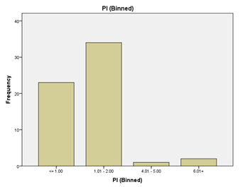
Graph 1
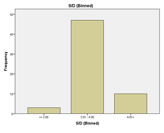
Graph 2
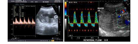
Figure 1: Image(A), Show the high S/D ratio with increased PI,RI at 34. 5 weeks. Image (B), Umbilical artery shows the reversal diastolic flow.
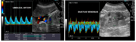
Figure 2: Image (A), High S/D ratio 3. 67 with thinning of diastolic flow. Image (b), shows the ductus venosus.
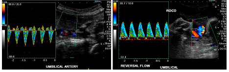
Figure 3: Image (A), shows the reversal of flow. Image (B), shows the absent and mild reversal of flow.
4. Discussion
In all intrauterine growth restricted fetuses with weight or Abdominal circumference less than 10th percentile for gestational age Umbilical artery Doppler should be performed [11]. Among Doppler parameters usually PI and S/D ratio are used and both are satisfactory to deal with intra-uterine growth restriction. When end-diastolic flow is absent than S/D cannot be measured and PI should be used [12]. The risk for perinatal morbidity and mortality is high in intra-uterine growth restricted fetuses [13]. The severity of umbilical artery flow abnormalities and gestational age determines how intra-uterine growth restriction starts and how it will proceed [14]. Fetuses with lateral placentas have a 4 times more risk for intra-uterine growth restriction as compared to anterior or posterior placentas [15]. In this study we found an inverse relation of Doppler indices with advancing gestational age of normal fetuses. This is similar to reported by studies of Gupta, et al. [16], Ganesh Acharya, et al. [17], Pharuhas Chanprapaph, et al. [18], Major Satyabrat Kumar, et al. [19], and Sachin Kumar Singh, et al. [20]. Shivani Singh, et al [21] reported high Doppler indices in intra-uterine growth restriction fetuses as compared to normal. In their study mean RI was 0. 75, 0. 81, 0. 70, 0. 77 and mean S/D was 3. 47, 3. 48, 3. 21, 3. 19 at 30, 33, 36 and 39 weeks respectively in intra-uterine growth restriction fetuses. In our study the RI was 0. 51-1. 00 and S/D ratio was 2. 01-4. 00 of IUGR fetuses at 30-39 weeks of gestation. Ranjan K. Sahoo, et al. , in India reported high Doppler indices in intra-uterine growth restriction group as compared to normal group [22]. In a study conducted by Merina Gayawali, et al. [24] in Nepal 72 out of 140 subjects suspected with intra-uterine growth restriction showed abnormal umbilical artery Doppler findings, 57 of these with S/D ratio greater than 324. This study is strongly related to our study that the S/D ratio was greater than 3 of 20 intra-uterine growth restriction fetuses. Sattar MA et al. , [25] found high values of Doppler PI, low Biophysical profile score and low AFI in intra-uterine growth restriction suspected fetuses group as compared to the normal group. Their findings of Doppler’s results are similar to our results. In their study the mean values of PI in intra-uterine growth restriction group were 1-1. 425. In my study the PI values of intra-uterine growth restriction fetuses lie between1. 0-2. 0. Azmat, et al. , showed 38 fetuses with absent end diastolic flow and 12 fetuses with reversed end diastolic flow out of 50 intra-uterine growth restriction fetuses [23]. Noha et al. , conducted a study on sixty high risks intra-uterine growth restricted fetuses and found absent end-diastolic flow in 10 fetuses and reversed end-diastolic flow in 8 fetuses. They also observed umbilical vein monophasic pulsation in 8 fetuses and triphasic flow in 2 fetuses [26]. In our study of 30 intra-uterine growth restricted fetuses we found 18 fetuses having decreased end diastolic flow and 12 fetuses having absent end diastolic flow. The results of our stud are comparable with studies [23, 26]. The results of our study confirmed the importance of Doppler ultrasound in fetuses with suspicion of intra-uterine growth restriction. Doppler can confirm whether a suspected intra-uterine growth restricted fetus is an actual case of intra-uterine growth restriction or a normal fetus, it will also describe the severity of intra-uterine growth restriction according to the status of end diastolic flow.
5. Conclusion
This study has provided evidence that Doppler indices (PI, RI, and S/D ratio) in intra-uterine growth restricted fetuses are directly proportional to gestational age while in normal fetuses they are inversely proportional. The findings of this study are similar to previous studies. These findings will help to reduce mortality and morbidity in IUGR suspected fetuses by timely diagnosis and management.
References
- Gilani SA, Javaid A, Bala AA. Doppler ultrasound assessment of the ductus venosus in the normal fetus between 20 and 40 weeks gestation in the Pakistani population. Ultrasound 18 (2010): 148-151.
- Fleischer A, Schulman H, Farmakides G, Bracero L, Blattner P, Randolph G. Umbilical artery velocity waveforms and intrauterine growth retardation. American Journal of Obstetrics & Gynecology 151 (1985): 502-505.
- Manning FA, Hohler C. Intrauterine growth retardation: diagnosis, prognostication, and management based on ultrasound methods. The principles and practice of ultrasonography in obstetrics and gynecology. 4th ed. Norwalk, Conn. : Appleton & Lange (1991): 331-348.
- Trudinger BJ, Stevens D, Connelly A, Hales JR, Alexander G, Bradley L, et al. Umbilical artery flow velocity waveforms and placental resistance: the effects of embolization of the umbilical circulation. American journal of obstetrics and gynecology 157 (1987): 1443-1448.
- Kingdom JC, Burrell SJ, Kaufmann P. Pathology and clinical implications of abnormal umbilical artery Doppler waveforms. Ultrasound in Obstetrics and Gynecology: The Official Journal of the International Society of Ultrasound in Obstetrics and Gynecology 9 (1997): 271-286.
- Bernstein IM, Horbar JD, Badger GJ, Ohlsson A, Golan A, Vermont Oxford Network. Morbidity and mortality among very-low-birth-weight neonates with intrauterine growth restriction. American journal of obstetrics and gynecology 182 (2000): 198-206.
- Mari G, Abuhamad A, Brumfield J, Ferguson JE. 669 Doppler ultrasonography of the middle cerebral artery peak systolic velocity in the fetus: Reproducibility of measurement. American Journal of Obstetrics & Gynecology 185 (2001): S261.
- Veille JC, Hanson R, Tatum K. Longitudinal quantitation of middle cerebral artery blood flow in normal human fetuses. American journal of obstetrics and gynecology 169 (1993): 1393-1398.
- Kozar M, Tonhajzerova I, Mestanik M, Matasova K, Zibolen M, Calkovska A, et al. Heart rate variability in healthy term newborns is related to delivery mode: a prospective observational study. BMC pregnancy and childbirth. 18 (2018): 264.
- Baschat A, Gembruch U, Weiner C, Harman C. 0325 Longitudinal Changes Of Arterial And Venous Doppler In Fetuses With Intrauterine Growth Restriction (iugr). American Journal of Obstetrics and Gynecology 184 (2001): S103.
- Lausman A, Kingdom J, Gagnon R, Basso M, Bos H, Crane J, et al. Intrauterine growth restriction: screening, diagnosis, and management. Journal of Obstetrics and Gynaecology Canada 35 (2013): 741-748.
- Berkley E, Chauhan SP, Abuhamad A, Society for Maternal-Fetal Medicine Publications Committee. Doppler assessment of the fetus with intrauterine growth restriction. American journal of obstetrics and gynecology 206 (2012): 300-308.
- Fatima B. Validity of Umbilical Artery Doppler Ultrasound in Diagnosis of Intrauterine Growth Restricted Fetuses. Journal of Rawalpindi Medical College 19 (2015): 140-142.
- Turan OM, Turan S, Gungor S, Berg C, Moyano D, Gembruch U, et al. Progression of Doppler abnormalities in intrauterine growth restriction. Ultrasound in Obstetrics and Gynecology: The Official Journal of the International Society of Ultrasound in Obstetrics and Gynecology 32 (2008): 160-167.
- ERDOLU MD, KÖ?Ü? A, KÖ?Ü? N, D?LMEN G, Kafali H. Relationship between placental localisation, birth weight, umbilical Doppler parameters, and foetal sex. Turkish journal of medical sciences 44 (2014): 1114-1117.
- Edan BJ, Al Esawi RW, Shukur ZY. Umbilical Artery Flow Velocity in Pregnancy Induced Hypertension at Third Trimester. Kufa Medical Journal 17 (2017): 92-100.
- Acharya G, Wilsgaard T, Berntsen GK, Maltau JM, Kiserud T. Reference ranges for serial measurements of umbilical artery Doppler indices in the second half of pregnancy. American journal of obstetrics and gynecology 192 (2005): 937-944.
- Chanprapaph P, Wanapitak C, Tongsong T. Umbilical artery Doppler waveform indices in normal pregnancies. Thai J Obstet Gynaecol 12 (2000): 103-107.
- Srikumar S, Debnath J, Ravikumar R, Bandhu HC, Maurya VK. Doppler indices of the umbilical and fetal middle cerebral artery at 18-40 weeks of normal gestation: A pilot study. Medical Journal Armed Forces India 73 (2017): 232-241.
- Philip B, Kurian S. ASSESSMENT OF FOETAL COMPROMISE BY DOPPLER ULTRASOUND INVESTIGATION OF THE FOETAL CIRCULATION. Journal of Evidence Based Medicine and Healthcare 4 (2017): 4364-4367.
- Singh S, Verma U, Shrivastava K, Khanduri S, Goel N, Zahra F, et al. Role of color doppler in the diagnosis of intra uterine growth restriction (IUGR). International Journal of Reproduction, Contraception, Obstetrics and Gynecology 2 (2016): 566-572.
- Ranjan K, Sahoo, Nayak S, Sitansu K, Panda, Pravkar B, et al. Doppler Assessment of the Fetus with Intrauterine Growth Restriction. Int. J. Pharm. Sci. Rev. Res 32 (2015): 162-170
- Ali A, Ara I, Sultana R, Akram F, Zaib MJ. COMPARISON OF PERINATAL OUTCOME OF GROWTH RESTRICTED FOETUSES WITH NORMAL AND ABNORMAL UMBILICAL ARTERY DOPPLER WAVEFORMS. Journal of Ayub Medical College Abbottabad 26 (2014): 344-348.
- Gyawali M, Sharma P, Lalchan S, Tiwari PK. Prediction of neonatal outcome by umbilical artery velocimetry in intrauterine growth restriction: a study in western Nepal. American Journal of Public Health Research 3 (2015): 139-143.
- Sattar MA, Wahed SR. Intrauterine growth restriction: role of ultrasound and color flow Doppler. AAMJ, (2011): 187-208.
- AbdelMaboud NM, Elsaid HH. Role of venous Doppler evaluation of intrauterine growth retardation. The Egyptian Journal of Radiology and Nuclear Medicine 46 (2015): 167-174.