Magdalena Filcek1*, Afaf El-Ansary2, Marzena Dominiak3, Mayank Vats4
1Specialist of Neuroarchitecture, Master of Architecture Interior Design at Academia of Fine Art, designer, researcher, PhD pending of Safety Engineering, Faculty of Security Studies, General Tadeusz Kościuszko, Military University of Land Forces Wroclaw, Poland
2Autism Research and Treatment Center, King Saud University, KSA
3Dental Surgery Department Wroclaw Medical University, Wrocław, Poland
4Pulmonology, Sleep Medicine, Interventional Pulmonologist and Critical Care Medicine, researcher, Visiting Prof. SMS
Hospital in India, Prof. Sheikh Mohammad Bin Rashid University, Dubai Medical College, Rashid Hospital, Dubai
Hospital, Dubai, UAE
*Corresponding Author: Magdalena Filcek, Specialist of Neuroarchitecture, Master of Architecture Interior Design at Academia of Fine Art, designer, researcher, PhD pending of Safety Engineering, Faculty of Security Studies, General Tadeusz Kościuszko, Military University of Land Forces Wroclaw, Poland.
Received: 06 May 2026; Accepted: 05 June 2026; Published: 30 July 2026
DOI: 10.26502/jppd.2572-519X0280
ShareAutism Spectrum Disorder (ASD), Attention-Deficit/Hyperactivity Disorder (ADHD) and Post-Traumatic Stress Disorder (PTSD) are complex neuropsychiatric conditions whose pathophysiology until now remains unclear. In this article, we expand on the author's earlier hypothesis that Mast Cell Activation Syndrome (MCAS) and impaired histamine metabolism, called Histamine Intolerance (HIT), may play a key role in the development of the commonly observed symptoms of this disease. By integrating current scientific evidence on the role of mast cells, histamine, neurotransmitters, and biological, chemical, physical, nutritional, and environmental factors, this study aims to elucidate potential mechanisms underlying the etiopathogenesis of these disorders. Systematic review and meta-analyses presenting a significant correlation between MCAS, histamine accumulation, GABA reduction, glutamate hyperactivity, and neuroinflammation, leading to excitotoxicity, neuronal damage and clinical manifestations. The author suggests that symptoms such as sensory hypersensitivity, gastrointestinal dysfunction, sleep disturbances, and emotional instability, commonly observed in patients with ASD, ADHD, PTSD, MCAS, and HIT, may result from histamine hyperactivity. Excess histamine leads also to activation of H1–H4 receptors, affecting multiple physiological systems, including the respiratory, gastrointestinal, circulatory, immune, and nervous systems. It can also cross the blood-brain barrier, promote blood clot formation, stimulate H3 receptors in the brain, and trigger neuroinflammation. As a working hypothesis of therapeutic interventions, it is proposed that a combination of sensory-soothing environments, internal rhythm–synchronizing technologies (such as Vinci Power Nap®), low-histamine diets, and targeted supplementation (vitamins D, A, B6, B9, B12, C, zinc, copper, H1–H4 receptor blockers, and DAO enzyme, etc.) may alleviate symptoms by modulating mast cell activity and excitotoxicity, improving sleep quality, and promoting balanced immune and nervous system responses, which was noticed in pilot experiments. These findings may inspire further research for randomized clinical trials (RCTs) to validate these strategies as well as for further research to explore the role of mast cells and histamine in neuroimmune disorders including also stroke, Alzheimer and Parkinson disease.
ASD, ADHD, PTSD, Mast cells, Histamine, Enzyme DAO, H1, H2, H3 receptors, GABA, Glutamate, Excitotoxicity, Neuroinflammation, Neuroarchitecture.
ASD articles; ADHD articles; PTSD articles; Mast cells articles; Histamine articles; Enzyme DAO articles; H1 articles; H2 articles; H3 receptors articles; GABA articles; Glutamate articles; Excitotoxicity articles; Neuroinflammation articles; Neuroarchitecture articles
1. Introduction
Autism Spectrum Disorder (ASD), Attention-Deficit/Hyperactivity Disorder (ADHD), and Post-Traumatic Stress Disorder (PTSD) are complex neuropsychiatric conditions that significantly affect the quality of life of patients and their families. According to the most recent estimates from the CDC’s Autism and Developmental Disabilities Monitoring (ADDM) Network, approximately 1 in 31 (3.2%) children aged 8 years were diagnosed with ASD in 2022 [1], [2], compared with approximately 1 in 59 children in 2020 [3]. A meta-analysis of multiple studies demonstrated that the global prevalence of ADHD among children and adolescents is approximately 8.0%, with rates of 10% in boys and 5% in girls in the analyzed epidemiological samples [4]. Between 2009 and 2017, based on parent-reported data, approximately 1 in 6 (17%) children aged 3–17 years in the United States were diagnosed with at least one developmental disability. These included, among others, ASD, ADHD, blindness, and cerebral palsy [6]. Post-traumatic stress disorder (PTSD) is a mental health condition that may develop following exposure to, or witnessing of, a traumatic event. Such events include, but are not limited to, armed conflict, natural disasters, terrorist attacks, motor vehicle accidents, as well as prolonged abuse or neglect during childhood [7]. Experts indicate that PTSD arises from stress-induced neurobiological changes in the brain. In many individuals, symptoms emerge shortly after the traumatic event, whereas in others they may develop after a prolonged latency period, sometimes years later [8]. PTSD affects approximately 6–8% of the general population across all racial, ethnic, and socioeconomic groups [5].
The aforementioned disorders are characterized by a complex etiology involving genetic, environmental, and immunological factors, and despite extensive research, their pathophysiological mechanisms remain incompletely understood, which continues to hinder the development of effective therapeutic strategies. Symptoms such as, among others, sensory hypersensitivity, gastrointestinal disturbances, allergic manifestations, cognitive difficulties, emotional instability, and sleep disorders suggest the presence of shared neurobiological mechanisms [9], [10]. Notably, these symptoms show a strong overlap with those described in the recently identified and published “histamine storm” mechanism implicated in the development of systemic inflammatory response syndrome (SIRS) and sepsis [11]. Recognition of these correlations led the author to further observations pointing to a potential role of excessive Mast Cell Activation Syndrome (MCAS) and Histamine Intolerance (HIT) as contributing factors to neuroinflammation and dysregulation of neurotransmission in ASD, ADHD, and PTSD [12]. Mast cells, localized predominantly in barrier tissues such as the skin, mucosal surfaces, gastrointestinal tract, lungs, heart, and also within the vascular endothelium, are highly sensitive to a wide range of environmental biological, chemical, and physical stimuli. These include, among others, extreme cold or heat, cold and dry air to breathe [13], mechanical pressure, vibration, ionizing radiation, sensory stressors, allergens, pharmaceuticals, toxins, and heavy metals. Upon activation, mast cells undergo degranulation and release a broad spectrum of pro-inflammatory mediators, most notably histamine. Histamine exerts its effects through activation of H1–H4 receptors, thereby amplifying local and systemic inflammatory responses and modulating neurotransmission, ultimately influencing central and peripheral nervous system function [14], [15], [16]. It is important to emphasize that MCAS represents a distinct clinical entity from mastocytosis, differing in pathophysiology, diagnostic criteria, and clinical course. In addition, the exogenous histamine load derived from dietary sources plays a significant role in overall histamine homeostasis. Impaired histamine metabolism, known as histamine intolerance (HIT)— most commonly associated with reduced activity or deficiency of diamine oxidase (DAO)—may further exacerbate spectrum of symptoms as well as neurological dysfunction. Excessive levels of this endogenous and exogenous biogenic amine, which also functions as a neurotransmitter, have been shown to critically contribute to the dysregulation of cognitive, emotional, and autonomic processes [17].
A growing number of studies indicate that mast cell activation syndrome (MCAS) and histamine intolerance (HIT) may be linked to alterations in neurotransmitter homeostasis, particularly affecting the balance between glutamate and gamma-aminobutyric acid (GABA). Glutamate, the principal excitatory neurotransmitter, and GABA, the primary inhibitory neurotransmitter, play critical roles in the regulation of neuronal activity and synaptic homeostasis [18]. Dysregulation of this excitatory–inhibitory balance may lead to neuronal hyperexcitability, altered sensory processing, hypersensitivity, and impairments in emotional regulation. Histamine released within the central nervous system (CNS) modulates neurotransmission through multiple receptor subtypes, including the H3 receptor, which functions predominantly as a presynaptic autoreceptor and heteroreceptor. Activation of H3 receptors inhibits the release of several neurotransmitters, notably GABA, thereby indirectly promoting increased glutamatergic signaling [19]. This shift toward excitatory dominance may contribute to heightened neuronal excitability, excitotoxicity, and the initiation or amplification of neuroinflammatory processes [20], [21], [22]. Building upon published evidence and observational data, the present review investigates these mechanisms, their possible involvement in the pathophysiology and symptom expression of ASD, ADHD, and PTSD, and the potential role of therapeutic approaches aimed at modulating mast cell function and histamine metabolism regulation. Such interventions include low-histamine dietary approaches, optimization of the external environment to minimize stressors, mast cell stabilization strategies (e.g., vitamin supplementation and antihistamine use), and reduction of histamine levels through enzymatic degradation via diamine oxidase (DAO) supplementation [23], [24], [11], [25], [26].
Hypothesis: overlapping factors that elevate histamine levels may act synergistically, thereby amplifying one another and contributing to a broad range of symptoms observed in ASD, ADHD, and PTSD:
- Environmental factors → Mast cell activation ↑ → histamine ↑ → H1, H2, H3, H4 receptors ↑ H3 → GABA ↓ → Excessive glutamatergic activity ↑ → neuronal excitability ↑ → Excitotoxicity and neuroinflammation ↑ → Oxidative stress ↑ → Symptom severity ↑
- High-histamine foods ↑ + low diamine oxidase (DAO) activity ↓ → Elevated histamine levels ↑↑ → H1, H2, H3, H4 receptors ↑ H3 → GABA ↓ → Excessive glutamatergic activity ↑ → neuronal excitability ↑ → Excitotoxicity and neuroinflammation ↑ → Oxidative stress ↑ → Symptom severity ↑
- Amplification (1+2): Environmental factors + High-histamine foods ↑ + low diamine oxidase (DAO) activity ↓ → histamine levels ↑↑↑ → H1, H2, H3, H4 receptors ↑↑ → GABA ↓↓ → Excessive glutamatergic activity ↑↑ → Increased neuronal excitability ↑↑ → Excitotoxicity and neuroinflammation ↑↑ → Oxidative stress ↑↑ → Mast cell activation ↑ → histamine levels ↑↑↑ → Exacerbated symptoms of ASD, ADHD, and PTSD ↑↑ (Picture 1)
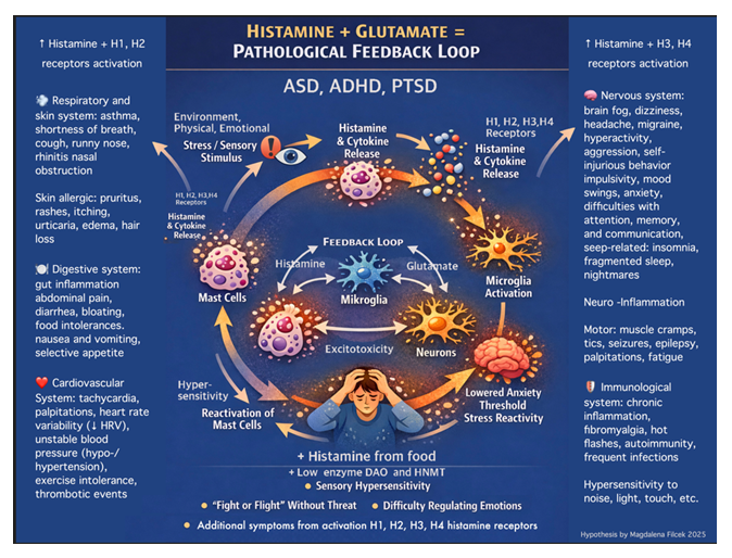
Picture 1: Hypothesis: overlapping factors that elevate histamine and glutamate levels, also activating H1-H4 receptors may act synergistically, thereby amplifying one another and contributing to a broad range of symptoms observed in ASD, ADHD, and PTSD. Illustration was generated using AI (ChatGPT 4.0.) and reviewed and completed by the author.
2. The Relationship Between MCAS, HIT, Histamine Accumulation, and ASD, ADHD, PTSD
Conclusions from meta-analyses: MCAS + HIT → Excess histamine → Activation of H3 receptors → Reduced GABA release → Excessive glutamatergic activity → Excitotoxicity and neuroinflammation → Disrupted homeostasis and neuronal damage → Potentially explains shared symptoms of ASD, ADHD, and PTSD.
An increasing body of evidence from meta-analyses demonstrates a significant association between immune-mediated mast cell activation (MCAS), including the degranulation of histamine, and dysregulated histamine metabolism (histamine intolerance, (HIT)), and neuroinflammation and the neuropsychiatric symptoms observed in ASD, ADHD, [27], [28], [29], [30], as well as in PTSD [31]. Some of the most comprehensive and detailed meta-analyses describing an overlooked mechanism helpful in understanding this connection is explained in the article titled: “Discovery of the Mechanism of COVID-19, SIRS and SEPSIS, Defense and Treatment. Mast Cells and Histamine Storm: An Overlooked Aspect in COVID-19 and in Ventilated Patients – Potential Role of Antihistamines” [11], also as one of the chapter in the book „Oddech” entitled: “Breathe with Vinci Power Nap® – Harmonize Your Body and Mind in the Sleep Cafe®: A Neuroarchitectural System for Synchronizing Sleep, Breathing, Heartbeat, and Brainwaves that Reduces Stress, Mast Cell Activation Stimuli, and Histamine” [27]. The author’s hypothesis identifies this overlooked mechanism as a potential pathophysiological factor underlying certain features of ASD, ADHD and PTSD [10], [27] because MCAS is associated with chronic histamine release, which exacerbates neuroinflammatory and behavioral symptoms also characteristic of HIT [32]. The analyzed studies indicates that patients with mastocytosis frequently experience neuropsychiatric symptoms, including depression (68–75%), anxiety, elevated stress levels or irritability (27–54%), cognitive impairment—particularly memory deficits (27–39%), headaches (55–69%), attention deficits and learning difficulties in 13% of children, dental [33] and neurodevelopmental disorders in (8–12%) of cases [34]. Several clinical features of MCAS and mastocytosis [35], including excessive histamine release, activation of histamine receptors (H1, H2, H3, H4), and reduced enzyme diamine oxidase (DAO) activity resulting in histamine intolerance (HIT), overlap with symptoms commonly observed in ASD, ADHD, and PTSD [24], [36], like:
- Gastrointestinal: abdominal pain, diarrhea, bloating, food intolerances, selective appetite.
- Skin allergic: pruritus, rashes, itching, urticaria, edema.
- Sensory hypersensitivity: light, sound, smell, and touch sensitivity.
- Neurological: headaches, migraines, “brain fog,” mood and emotional fluctuations.
- Respiratory: asthma, shortness of breath, cough,
- Cardiovascular: tachycardia, palpitations, heart rate variability (↓ HRV), unstable blood pressure (hypo- /hypertension), exercise intolerance, dizziness, thrombotic events, hypotension.
- Behavioral: hyperactivity, impulsivity, anxiety, self-injurious behavior, aggression, reduced social engagement.
- Motor: muscle cramps, tics, seizures, palpitations, fatigue.
- Cognitive: difficulties with attention, memory, and communication.
- Immunological: chronic inflammation, fibromyalgia, hot flashes.
- Neuronal: glutamatergic excitotoxicity, neuroinflammation, neuronal damage.
- Sleep-related: insomnia, fragmented sleep, nightmares.
These findings suggest a potential coexistence of MCAS and HIT, mediated through impaired histamine metabolism due to reduced activity of enzymes: DAO or histamine N-methyltransferase (HNMT). Such impairments have also been observed in patients with ADHD [37], ASD [38], as well as in individuals carrying polymorphisms in the AOC1 and HNMT genes [39]. These alterations can lead to histamine accumulation and potentially exacerbate the aforementioned symptoms, including neurological and behavioral manifestations. For the diagnosis of HIT, consumption of histamine-rich foods must coincide with DAO deficiency; otherwise, the condition may be more accurately classified as histamine intoxication [29]. The role of mast cells in brain inflammation and autism spectrum disorder has also been highlighted in expert discussions and presentations [40]. The author notes that very similar symptoms are also observed in PTSD (Mental Health America. What are the symptoms of PTSD?) [8], suggesting that experienced trauma and intense stress may have activated and sensitized mast cells [41], potentially leading to MCAS, HIT, and/or reduced DAO enzyme production. This hypothesis is promising but warrants further investigation in larger studies.
3. Localization and Biology of Mast Cells: Their Role in the Regulation of Inflammation and Neurotransmission
Mast cells (MCs), discovered 140 years ago by Paul Ehrlich, are tissue-resident cells found in various organs [42]. Mast cells originate in the bone marrow and circulate in the blood as immature precursors. They complete their differentiation only after homing to target tissues, typically localizing in perivascular regions on the abluminal side of the vascular endothelium [43]. This strategic positioning allows them to interact with other cell types, the extracellular matrix, and the vasculature. Mast cells are also capable of migrating from the blood into the brain under both physiological and pathological conditions [44].
Mast cells play a key role in host defense, acting as one of the first responders of the immune system and initiating immune responses—particularly at sites of contact with the external environment, such as the skin, mucous membranes (e.g., nasal mucosa), respiratory tract (e.g., lungs), and gastrointestinal tract (e.g., intestines). They are located in close proximity to peripheral nerve endings in various tissues, as well as in the heart and brain. Within the brain, mast cells are found in multiple regions, including: near the blood–brain barrier (in areas of increased permeability, suggesting a potential role as sentinels in pathogen responses) [45], the choroid plexus and the protective membranes of the brain and spinal cord, the layers that surround and protect the central nervous system (the meninges) [23], [24]. They are also present in the hypothalamus, where they may influence emotional regulation, appetite, and stress responses, suggesting a potential modulatory role on the HPA axis (hypothalamus–pituitary–adrenal) [46]. Stress, infection, autoimmune diseases, or neuroinflammatory conditions, may promote mast cell activation and recruitment within the central nervous system [25], showing the relationship and involvement of mast cells in the brain, including multiple sclerosis [47].
3.1 Mechanisms of Mast Cell Activation
Mast cells are key first responders to most harmful or extreme stimuli (stressors), i.e., those that could potentially compromise the biological integrity of the organism, providing immediate defense, stress response, repair, and survival mechanisms. Their role as the “first link in the immune response chain” is underscored by their strategic localization in tissues near blood vessels, nerve endings, and mucosal surfaces. Mast cells express receptors (e.g., FcεRI for IgE) that allow for rapid recognition of pathogens or allergens. Immune activation can occur through IgE-dependent mechanisms or independently of IgE, as mast cells are also sensitive to external stressors, including biological, chemical, and physical factors, as well as internal stimuli such as stress, emotions, and hormones [48].
- IgE-mediated triggers: seemingly harmless antigens, including many present in foods (e.g., proteins, eggs, nuts, wheat), medications (e.g., penicillin), inhalants (e.g., grass and tree pollens, house dust mites, animal dander), and contact allergens (e.g., latex, detergents, cosmetics, preservatives) can elicit IgE-mediated MCs activation [49].
- Biological triggers:
- Infections (viral, bacterial, parasitic), including COVID-19, Epstein-Barr virus (EBV, mononucleosis), and Lyme disease, can disrupt immune homeostasis and dysregulate mast cells [42], [50].
- Dental caries, pulpitis, periapical periodontitis, periodontal disease, peri-implantitis, and other chronic oral infections [33].
- Fungal exposure and mold infections can also act as biological triggers [51].
- Chemical triggers:
- Toxins (e.g., fire ant venom, hymenoptera toxins, mycotoxins), poisons, opioids, alcohol, and certain odors can directly or indirectly activate mast cells [52].
- Drug-induced triggers: Nonsteroidal anti-inflammatory drugs (NSAIDs, e.g., ibuprofen) [53], opioids [54], certain antibiotics (e.g., vancomycin) [55], anesthetics [56], [57], [58] and contrast agents can induce mast cell degranulation [59], [60]. Personalized approach is recommended to address drugs inducing MCs activation [61].
- Food allergens and intolerances: Gluten, dairy, histamine, biogenic amines, and sulfites can also stimulate mast cells via non-IgE mechanisms [62].
- Environmental and toxic exposures: Heavy metals (e.g., aluminum, mercury) [48], [63], some dental materials (acrylic, metal alloys (g., nickel-containing fillings), cements [33], orthopedic and dental implants constructed from metal alloys or IV- or V-generation titanium are subject to corrosion and a biological reaction known as tribobiocorrosion [63], [64], microplastic [65], [66], mold, and pesticides often provoke chronic inflammation and can “prime” the immune system for increased mast cells reactivity [67], [68].
- In same cases, vaccination can trigger immune reactions ranging from mild local inflammation to allergic responses. Certain vaccine components, such as adjuvants containing aluminum salts, may transiently interact with the immune system to enhance antigen presentation. In individuals with pre-existing hypersensitivity conditions, immune responses may be more pronounced, [68], [69], [70]. There is a distinction between oral ingestion of heavy metals and direct intravenous exposure [71] in relation to ASD, discussed in media commentary by Robert F. Kennedy Jr., Secretary of the U.S. Department of Health and Human Services, among others [72], [73], [74], [75].
- Physical triggers:
- Temperature changes excessive heat (including sunlight) or cold, dry and cold air to breathe, intense light, noise (including constant background noise from AC units, devices, or servers), pressure or constriction (e.g., shoes, elastic bands in socks or underwear), physical exertion (intense exercise, physical urticaria) can all activate MCs [76].
- Mechanical factors in the oral cavity can also play a significant role. For example, chronic pressure or irritation caused by poorly fitting dentures, dental crowns, orthodontic appliances, occlusal overload, or persistent soft tissue trauma can trigger sterile inflammation and neurogenic signaling (including the release of neuropeptides, particularly substance P and calcitonin gene-related peptide (CGRP) [33].
- Electromagnetic fields, radiation, ionizing radiation [77], and vibrations [78], [79], [80].
- Stress and internal factors:
- Physical or psychological stress, anxiety, trauma, and fatigue can activate mast cells [81], [67]. Acute short-term stress may transiently suppress mast cell degranulation via epinephrine and glucocorticoid cortisol, but chronic stress destabilizes this system, increasing mast cell numbers and reactivity [82]. Animal models demonstrate increased mast cell density and activity in the skin and gut under chronic stress, while in humans, chronic stress exacerbates allergic and gastrointestinal conditions (e.g., urticaria, IBS) in which histamine plays a key role [83].
- Oxidative stress and neurohormonal triggers: Certain neurotransmitters and neuropeptides—including Substance P, Compound 48/80 [46], [84], adrenomedullin, bradykinin, Calcitonin Gene-Related Peptide (CGRP), Corticotropin-Releasing Hormone / Factor (CRH/CRF), enkephalins/endorphins, 17β-estradiol, HK-1, Mastoparan, Nerve Growth Factor (NGF), Neurotensin (NT), Parathyroid Hormone (PTH), Stem Cell Factor (SCF), Vasoactive Intestinal Peptide (VIP), urocortin, IL-1, and IL-33—can stimulate mast cells [81], [84].
- Hormonal triggers: Hormonal fluctuations, including the menstrual cycle, pregnancy, and menopause, can influence mast cell activity. Estradiol, for example, can stimulate mast cell degranulation [85].
- Gastrointestinal triggers: The gut contains a high density of mast cells, making it a major site for activation. Conditions such as SIBO, dysbiosis, or leaky gut can excessively activate mast cells [86], [87].
- Impaired histamine metabolism (e.g., DAO deficiency) and elevated biogenic amines (tyramine, putrescine) may enhance mast cell activation by reducing histamine degradation, increasing gut permeability, and amplifying inflammatory signals or pseudoallergic reactions [88], [89].
- Dietary triggers: Additives, preservatives, and colorants in processed foods can provoke pseudoallergic mast cell reactions. Elimination diets that exclude gluten, lactose, casein, or caffeine are commonly used in individuals with ASD [90]. Increasing attention is also given to low-histamine diets (avoiding aged cheeses, fish, tomatoes, fermented foods) in children with impaired DAO/HNMT activity.
Mediator release by mast cells occurs very rapidly, whereas prolonged activation leads to de novo synthesis of mediators [91]. In many situations, degranulation does not result in mast cell death; these are resilient and multifunctional cells capable of surviving activation and responding repeatedly to subsequent stimuli [92]. Mast cell degranulation releases, among other mediators, histamine, cytokines (e.g., TNF-α, IL-6), and additional pro-inflammatory mediators, thereby affecting the immune, gastrointestinal, respiratory, and nervous systems, as well as neurological functions such as memory and stress responses [93]. Consequently, mast cells play a crucial role in regulating inflammation and neurotransmission, i.e., neuroimmune communication. They act as sentinels of immunity, but when overactive, they can be harmful [49].
Often, it is not a single factor but a combination of the elements described above that leads to the development of Mast Cell Activation Syndrome (MCAS). For instance, in individuals with a genetic predisposition, a viral infection combined with stress and food intolerance may together “overload the system,” potentially triggering even anaphylactic shock [49]. The first-line treatment for anaphylaxis is epinephrine (adrenaline), which stabilizes mast cell membranes and rapidly inhibits further mediator release [94]. Cortisol, which rises during stress, exerts immunosuppressive effects by reducing mast cell activity, inhibiting pro-inflammatory cytokine production, and limiting enzyme release. Synthetic glucocorticoids, analogs of cortisol, are used to treat allergic diseases precisely because they block mast cell activation [95]. However, chronic stress (prolonged elevation of cortisol and adrenaline) can gradually dysregulate the immune system. In some individuals under chronic stress, mast cells become hyperreactive or increase in number, which may exacerbate allergic reactions [96].
- Acute stress → epinephrine and cortisol → inhibition of mast cells (reduced histamine release).
- Chronic stress → potential dysregulation of the system and secondary mast cell hyperreactivity → increased histamine release.
3.2 Chronic Stress and KIT Mutation, Low-grade Inflammation
Errors in DNA repair or prolonged exposure to oxidative stress can lead to a KIT D816V mutation in exon 17 of the KIT gene in mast cells [97]. This mutation results in constitutive, SCF (Stem Cell Factor)-independent activation of the receptor, leading to excessive proliferation and activation of mast cells [98]. The KIT D816V mutation is characteristic of systemic mastocytosis (SM), which involves increased mast cell numbers in multiple tissues and can induce symptoms arising from excessive mediator release (e.g., histamine), similar to those observed in MCAS (mast cell hyperreactivity). The benefits of localized mast cell activation during infection contrast sharply with the detrimental effects of systemic activation, which reduces survival in sepsis induced by cecal ligation and puncture (CLP) [99]. Studies show that systemic mast cell degranulation significantly worsens survival during sepsis [100].
Chronic stress → KIT mutation → continuous mast cell proliferation + lowered activation threshold → easier mediator release → clinical manifestations of MCAS/mastocytosis → some overlapping symptoms of ASD [25], [30].
This explains why mast cells can become more sensitive and active with age, while mechanisms controlling inflammation weaken. Studies show an age-dependent increase in mast cell numbers in the gut and brain [101], accompanied by chronic low-grade inflammation, also referred to as “inflammaging.” The so-called “low-grade inflammation of histamine” is a chronic [102], mild inflammatory state in which histamine is continuously released by mast cells, most often in adipose tissue or the lungs, for example in obesity, chronic metabolic diseases, or allergies [103]. It does not produce dramatic symptoms of acute inflammation but contributes to long-term health problems.
3.3 Mast Cells, Meta-Inflammation and Obesity in ASD
Obesity is now widely viewed as a long-term inflammatory and immunometabolic condition linked to greater mast cell activity and a higher density of these cells in adipose tissue [102], along with increased histamine release [104], oxidative stress and endothelial dysfunction [105], as well as adipose tissue–driven “meta-inflammation” mediated by pro-inflammatory cytokines and adipokines such as leptin [106], [107]. These processes may contribute to asthma exacerbation, insulin resistance, hypertension, atherosclerotic changes, gastrointestinal inflammation [87], increased stroke risk [108], [101], and broader metabolic, cardiovascular, and immunological complications [109]. Histamine signaling further influences appetite regulation and fat metabolism through central H1/H3 (in brain) receptor pathways affecting decrease satiety at the same time increase appetite and have influence on energy expenditure [110], as well as peripheral H2 (in stomach and adipose tissue) receptor pathways involved in lipolysis (fat breakdown) and metabolic regulation like: gastric juice production and insulin secretion [111]. The relationship between obesity and histamine is well known and studied [112], [113]. Experimental studies indicate that pharmacological stabilization or inhibition of mast cells can reduce inflammation of adipose tissue and improve metabolism of glucose [114], supporting the concept that mast cell-targeted interventions may represent a promising therapeutic strategy in obesity-associated inflammatory and metabolic disorders [115].
Increased rates of overweight and obesity have been observed in children with ASD, potentially reflecting complex interactions between neuroinflammation, altered stress responses, sensory-related eating behaviors, gastrointestinal dysfunction, and metabolic dysregulation. Emerging evidence suggests that chronic mast cell activation and histamine-mediated inflammatory pathways may also contribute to these metabolic abnormalities [116], [117], [118], [119]. Mast cells are anatomically and functionally coupled to sensory nerves, autonomic nerves, enteric nerves, meningeal structures, vagus-nerve signaling, and brain-associated neuroimmune niches. They respond to neuropeptides, neurotransmitters, glutamatergic signals, stress mediators, and neural injury signals, while neurons and glial cells respond to mast cell-derived histamine, tryptase, cytokines, prostaglandins, leukotrienes, and extracellular vesicle cargo. Mast cells have therefore been conceptualized as signal converters between tissues and neurons [120]. Peripheral inflammation can also activate brain mast cells through vagus-nerve-dependent mechanisms involving glutamate and NR2B receptorsignaling [121], [122].
On the other hand peripheral inflammation can occur in the course of abdominal and visceral obesity. Of particular interest is the metainflammation that develops in the course of insulin resistance. As a result of the accumulation and subsequent hypertrophy of adipose tissue, inflammation develops. Cytokines (TNF-alpha, IL-6, MCP-1) are released, which leads to the activation of M1 macrophages. Cytokines block insulin signaling. This leads to increased release of free fatty acids and the development of hyperinsulinemia. This, in turn, causes lipogenesis and adipocyte hypertrophy. As a result, inhibition of lipolysis is impaired, leading to the development of systemic and organic insulin resistance. This results in the development of oxidative stress, leading to the development of numerous complications, including fatty degeneration of the bone marrow, which leads to decreased osteogenesis and increased lipogenesis (yellow bone). Mechanistically, the bone marrow functions as both a developmental origin and a target compartment for the organization of mast cells. Mast cells originate from hematopoietic stem and progenitor cells, with initial differentiation occurring within the bone marrow prior to progenitors entering the circulatory system and achieving full maturation in peripheral tissues. According to [123] adult mouse bone marrow contains mast cell progenitors capable of differentiating into mature mast cells, thus establishing a well-defined hematopoietic foundation for the replenishment and distribution of mast cells within tissues. The marrow niche is not merely a production site. It encompasses vascular sinusoids, endothelial cells, mesenchymal stromal cells, osteoblast-lineage cells, osteoclast-lineage cells, macrophages, adipocytes, extracellular matrix, cytokine gradients, and hematopoietic stem and progenitor cells, any of which may influence mast cell progenitor survival, migration, and maturation [124]. In cases of mast cell proliferative disease, marrow infiltration further exemplifies that mast cells can become integrated within the niche, thereby affecting bone, stromal, and hematopoietic processes [125].
In a physiological context, this axis links mast cell biology to the supply of progenitors, the competence of the marrow niche, tissue repopulation, clonalexpansion, skeletal turnover, and the remodeling of marrow adipose tissue. From a modeling perspective, bone-marrow coupling requires variables for progenitor abundance, progenitor egress, stromal support within the marrow, sinusoidal trafficking, marrow adiposity, clonal mast cell burden, and feedback mechanisms from peripheral tissue demand to marrow output. Tissue injury activates mast cells and neurons in parallel; neural mediators further stimulate mast cells; mast cell mediators sensitize sensory endings, autonomic circuits, enteric nerves, meningeal structures, and brain-associated neuroimmune compartments; the resulting pain, itch, nausea, vascular reflex, thermoregulatory response, cognitive change, or autonomic output feeds back onto tissue physiology and inflammatory behavior. This is a bidirectional tissue-neural-brain amplification loop rather than a local sensory annotation from the American Association of Neurological Surgeons (AANS), American Society of Neuroradiology (ASNR), Cardiovascular and Interventional Radiology Society of Europe (CIRSE), Congress of Neurological Surgeons (CNS), European Society of Neuroradiology (ESNR), European Society of Minimally Invasive Neurological Therapy (ESMINT), European Stroke Organization (ESO), Canadian Interventional Radiology Association (CIRA), Society of NeuroInterventional Surgery (SNIS), Society of Interventional Radiology (SIR), Society for Cardiovascular Angiography and Interventions (SCAI), and World Stroke Organization (WSO); [126].
3.4 Mast Cells in the Brain and Neuroinflammation
Mast cells in the brain are a growing focus of interest in neuroimmunology [127]. Their presence is particularly studied in the context of neurodegenerative diseases such as Alzheimer’s disease [128] and autism spectrum disorder (ASD) [30], where they may contribute to the development of neuroinflammation, in line with immunological hypotheses of these conditions. Their significant role has also been observed in multiple sclerosis and migraine [45], and understanding the pathophysiology of this phenomenon suggests that mast cell-stabilizing agents could be a promising therapeutic strategy for migraine [84]. A large body of research indicates that inflammatory processes in the brain play a key role in the development of neuropsychiatric disorders. During stress, the hypothalamus releases corticotropin-releasing factor (CRF), which, together with neurotensin (NT), can activate mast cells present in the brain. In response, these cells release inflammatory (e.g., histamine) and neurotoxic mediators, leading to blood-brain barrier (BBB) disruption, microglial activation, and the formation of inflammatory foci [129]. Consequently, mast cells may contribute to symptoms such as “brain fog,” headaches, or ASD-related behavioral and cognitive disturbances, which can be exacerbated by stress [23], [24].
Children with ASD often display difficulty regulating anxiety and exhibit exaggerated responses to subtle triggers. Environmental stressors can activate mast cells, which then influence microglia, resulting in abnormal synaptic pruning and disrupted neuronal communication [25], [48], [67]. Notably, children with ASD have been found to have significantly higher serum levels of CRF and NT compared to controls, further supporting their involvement in autism pathogenesis [12], [26]. Research indicates that carbon dioxide [130] and vitamin D3 can inhibit mast cell degranulation and histamine release, offering promising prospects for individuals with MCAS [131], [132]. Findings from Professor Afaf El-Ansary’s research, which show the beneficial effects of vitamin D3 supplementation in individuals with ASD, further support the hypothesis that overactive mast cells and the histamine they release may play a significant role in the development of autism symptoms, influencing neuroinflammatory processes in the brain [133], [10].
3.5 Mast Cells, Glial Cells, and Neuroinflammation: Shared Mechanisms
Mast cells belong to the effector cells of the innate immune system, and it is they—not microglia—that act as the “first responders” to injury or activating stimuli [134]. Mast cells can serve a protective role, responding immediately and limiting cellular disturbances and brain damage by releasing both cytotoxic substances and factors that support regeneration. However, their chronic activation carries the risk of severe, destructive consequences [135]. Allergens, neuropeptides, stress, or toxins can activate mast cells to degranulate, releasing mediators such as histamine, which leads to blood–brain barrier (BBB) disruption and microglial activation, ultimately promoting neuroinflammation [29]. Glial cells, including microglia, produce pro-inflammatory substances that strongly influence central nervous system function. They modulate neuropathic pain, seizure activity, and neuronal damage. Glial cells communicate with other non-neuronal cells, primarily mast cells, and respond to their pro-inflammatory signals, which can exacerbate symptoms of neurodegenerative diseases, accelerate disease progression, and increase pain perception.
Astrocytes involved in chronic inflammatory disorders, including multiple sclerosis and ASD, are often found near mast cells, sharing the perivascular space [136], [137]. Both mast cells and glial cells can initiate endogenous regulatory mechanisms in response to chronic inflammation [138]. Consequently, CNS neurons may be “attacked” by a network of microglia–astrocytes–mast cells, particularly when regulation of these non-neuronal cells (e.g., mast cells) is insufficient due to excessive or chronic exogenous and/or endogenous stimuli and limited capacity to suppress their activity [138]. In neuroinflammatory disorders, the immune response can destructively affect nervous system cells and tissues following Karolinska Institute [139]. Increasing evidence points to interactions between mast cells and glial cells, offering new opportunities for therapies targeting neuroinflammation. Such strategies could involve selective modulation of mast cell activity, which plays a role in regulating neuronal hypersensitivity in both the peripheral and central nervous systems [140].
4. Histamine as a Hormone/Neurotransmitter – a Mediator of Coagulation, Interacting with H1–H4 Receptors in the Immune, Respiratory, Digestive, and Nervous Systems
Histamine is an organic compound belonging to the biogenic amine group and plays a key role in human physiology. It was first chemically characterized in 1910 by George Barger and Henry H. Dale during their studies of ergot alkaloids, where its pharmacological properties were first described [141]. Dale's subsequent research in the 1920s demonstrated the profound physiological effects of histamine, particularly its role in smooth muscle contraction, vasodilation, regulation of blood pressure, and stimulation of gastric acid secretion, leading to the concept of histamine as a biologically active mediator in the body [142], [143]. Later work by Ash and Schild contributed to further understanding of the pharmacology of histamine receptors, including the distinction of H1 receptor-mediated effects and the development of the antihistamine theory [144]. Histamine has proven to be crucial in allergies, anaphylactic reactions, stomach ulcers, and chronic inflammation. “The histaminergic system modulates various processes, including wakefulness, feeding, learning, and memory consolidation.” [145]. It is a biologically active substance, often classified as a tissue hormone or neurohormone, because it regulates multiple physiological processes and systems. Chemically, histamine is 2-(1H-imidazol-4-yl) ethylamine [146], produced from the amino acid histidine through the action of the enzyme histidine decarboxylase (HDC) [147]. Structurally, histamine consists of an imidazole ring attached to an ethylamine chain, with the molecular formula C₅H₉N₃. Histamine functions as a signaling molecule in intercellular communication and is recognized as one of the main mediators of allergic reactions and inflammatory processes [148].
4.1 Sources of Histamine in the Body
- Endogenous production :
- Histamine is produced endogenously, mainly by immune system cells such as mast cells and basophils, and it is also synthesized by macrophages. It is stored in an inactive form within the granules of mast cells and basophils until various stimuli trigger its release, such as allergens, strong sensory or environmental stimuli, injuries, or infections [149].
- In the central nervous system, histamine is produced by histaminergic neurons as well as perivascular mast cells, while the brain endothelium also serves as a physiological reservoir for histamine [150].
- Certain gut bacteria, including strains from the genera Lactobacillus (e.g., L. reuteri), Enterococcus, Morganella morganii, and Klebsiella, can also produce histamine in the intestines, especially in the presence of a high-histidine diet. This microbial histamine production can irritate the intestinal mucosa, causing diarrhea and abdominal pain [151], [147].
- Histamine is produced when the amino acid histidine undergoes decarboxylation, a reaction driven by the bacterial enzyme named histidine decarboxylase (HDC). In the intestinal microbiota, this process may lead to excessive histamine formation, which in some cases is associated with symptoms of histamine intolerance (HIT) [147]. This pathway can activate immune responses, including inflammation and allergy-like effects, as well as affect the gastrointestinal system. After entering the bloodstream, elevated histamine levels may trigger reactions similar to HIT—such as headaches, skin flushing, and pruritus—often referred to as pseudoallergic responses [152], [153].
- Factors that increase endogenous histamine production:
- Some medications can strongly inhibit the activity of the DAO enzyme, which may lead to the development of HIT. Such medications include aspirin (acetylsalicylic acid), nonsteroidal anti-inflammatory drugs (NSAIDs), morphine, clavulanic acid, acetylcysteine, cimetidine, and isoniazid [154].
- Since many of these medications are administered to children for various illnesses [155], [156], it is also important to consider the possibility of DAO inhibition in young children, especially when medications are used long-term or for chronic conditions. However, there is currently no conclusive evidence confirming this effect. Nevertheless, some of these medications are excluded in low-histamine diets for children [157].
- A diet rich in histidine
- Imbalances in gut microbiota (dysbiosis)
- Certain gastrointestinal disorders (e.g., SIBO–small intestinal bacterial overgrowth) [152].
- Exogenous sources:
- Histamine is also supplied through food, especially fermented, pickled, and moldy products, or those that undergo bacterial fermentation (e.g., improperly stored fish). Excess dietary histamine can lead to food poisoning [152], [158]. Foods and beverages high in histamine include:
- Yeast and yeast extracts
- Aged cheeses such as camembert, parmesan, and blue cheeses
- Fermented dairy products
- Alcohol, especially red wine and beer, and soy sauce
- Canned fish, such as mackerel and tuna, and other canned products
- Sauerkraut, pickles, and other fermented vegetables
- Smoked products, such as sausage, ham, bacon, and salami
- Seafood and shellfish, edible insects
- Chocolate and nuts
- Carrageenan, monosodium glutamate (MSG)
- Certain fruits: strawberries, citrus, pineapple, ripe bananas, raspberries, kiwi
- Certain vegetables: tomatoes, avocado, eggplant, spinach
- Vinegar and vinegar-containing products: ketchup [159], mayonnaise, mustard, pickles [160], [161], [162], [163].
- Foods containing other biogenic amines, such as tyramine or putrescine, which often occur alongside histamine, can competitively inhibit the activity of the DAO enzyme. As a result, even normally tolerated amounts of histamine may trigger symptoms of HIT [154], [164]. Examples of such foods include fish, fermented sausages, and sauerkraut [165], [166].
- Histamine is also supplied through food, especially fermented, pickled, and moldy products, or those that undergo bacterial fermentation (e.g., improperly stored fish). Excess dietary histamine can lead to food poisoning [152], [158]. Foods and beverages high in histamine include:
Histamine released from mast cells, as well as that obtained from food, accumulates in the body, which can lead to exceeding its optimal levels and intensifying its effects, especially when the DAO enzyme, responsible for its breakdown, is at low levels. This situation results in so-called “histamine excess” in the body, which can have clinical relevance in certain inflammatory conditions or in HIT. To diagnose HIT, the consumption of histamine-rich foods must be accompanied by a DAO deficiency [164]. Otherwise, the condition should be diagnosed as histamine poisoning [153].
4.2 Functions of Histamine – Positive and Negative Effects
Histamine, in appropriate amounts, is essential for proper bodily function and affects multiple systems [154]:
- Immune and inflammatory system: Acts as a mediator of immune responses and allergic reactions, releasing other pro-inflammatory substances via its receptors [148], supports the body’s “first-line defense” against bacteria, viruses, parasites and other enemies [167].
- Nervous system: Functions as a neurotransmitter; through H1 and H2 receptors, it regulates the sleep-wake cycle (e.g., promoting wakefulness) [9], [168] and stress [169]. By modulating microglial function, histamine indirectly affects neuronal survival and functions such as memory [170] and cognitive processes [171]. In the brain, it acts on H3 receptors, modulating release of other neurotransmitters and inflammatory states [172], [20], [21].
- Digestive system: Stimulates gastric acid production, aiding digestion [173].
- Respiratory system: Involved in the pathogenesis of bronchial asthma [174], [175].
- Circulatory system: Regulates cardiovascular function and promotes blood coagulation [176], [177], [178], [179]. Histamine has a direct and significant effect on the heart, as H1 and H2 receptors are physiologically present in the myocardium, conduction system, and coronary vessels, affecting heart rate, blood pressure, and coronary contraction [180], [181].
- Other roles: Influences fetal development, smooth muscle contraction (intestine, uterus, bronchi), endometrium [182], menstrual pain [183], thermoregulation, appetite control, and mediates anaphylactic reactions [168], [184], [185], [186], [187], [188].
- Wound healing and tissue repair: Histamine increases vascular permeability to allow immune cells and nutrients to reach damaged tissue. Can promote angiogenesis and tissue remodeling [189], [190], [191], [192].
However, in excessive amounts histamine is harmful and can lead to frequent, sometimes health-threatening multisystem symptoms. Elevated histamine levels may contribute indirectly to the activation of the coagulation cascade, which can increase the risk of thrombus formation and disseminated intravascular coagulation (DIC). As a consequence, this may be associated with higher blood pressure and serious cardiovascular complications such as stroke, myocardial infarction, and tachycardia. At the same time, histamine excess can increase vascular permeability, which may result in hypotension, edema, and urticaria, as well as greater sensitivity to pain. It is also linked with a range of allergic-type manifestations, including pruritus, skin rashes, sneezing, watery rhinorrhea, and asthma. In the gastrointestinal tract, high histamine levels may lead to hyperacidity of the stomach, gastroesophageal reflux, nausea, vomiting, and diarrhea. Other possible systemic effects include insomnia, chronic fatigue, muscle cramps, and hair loss, as well as a tendency toward chronic neuroinflammatory states. In addition, associations have been reported with obesity, rheumatoid arthritis, and various chronic inflammatory conditions.
This suggests that excess histamine in the body may exacerbate symptoms of ASD, ADHD, and PTSD. At the same time, exhaustion of coagulation factors and platelets increases the risk of bleeding, which may occur in sepsis, severe infections, malignancies, pregnancy complications, and similar conditions [12], [25], [26]. Similar symptoms were observed in a VAERS-based study from 2005 following administration of a combined tetanus-containing vaccine with hepatitis B vaccination [69], [70]. These findings suggest the possibility that certain components, for example, may chronically stimulate mast cells to release histamine.
After activation (degranulation), mast cells release inflammatory mediators such as tryptase, heparin, cytokines, and histamine. Histamine not only performs the functions described above, but also “calls for reinforcements” through histamine receptors H1, H2, H3, and H4, which may lead to the release of cytokines (e.g., TNF-α, IL-1, IL-4, IL-6, IL-8, IL-10), thereby initiating and shaping the entire immune response in both allergic reactions, infections, and tissue repair processes. If this process becomes amplified, inflammatory reactions within tissues may activate additional mast cells, leading to the release of further amounts of histamine. This can contribute to a vicious cycle known as a “histamine storm,” occurring in organs such as the lungs, intestines, heart, and brain, and may result in increased thrombus formation and multi-organ inflammation—a mechanism that can lead to systemic inflammatory response syndrome (SIRS) and, in the most severe cases, to sepsis [11], [193], [194].
4.3 Histamine Receptors H1, H2, H3, and H4 on Mast Cells and on Histaminergic Neurons
Histamine functions both as a hormone and a neurotransmitter. It influences the permeability of the blood–brain barrier (BBB) and exerts its effects through four types of receptors (H1–H4) located in various tissues, including the brain [195]. This widespread distribution explains the pleiotropic effects of histamine, ranging from allergic and neurological responses to regulatory functions [154].
- The H1 receptor is expressed, among others, in vascular endothelium, the lungs and cardiac muscle, smooth muscle (including bronchi, gastrointestinal tract, and uterus), the brain, neutrophils, and macrophages. Upon activation by histamine, H1 receptors contribute to responses such as smooth muscle contraction (e.g., bronchoconstriction leading to asthma symptoms and uterine contractions during menstruation), as well as relaxation of vascular smooth muscle, resulting in vasodilation and increased vascular permeability (edema, erythema). H1 receptors play a key role in allergic reactions and are also involved in myocardial inflammation. In the central nervous system, H1 receptors are predominantly postsynaptic and mediate the excitatory effects of histamine, including wakefulness and stress responses [196], [180].
- The H2 receptor is expressed in the stomach and cardiac muscle, as well as in T lymphocytes and basophils. Upon activation by histamine, it stimulates gastric acid secretion, contributes to the regulation of cardiac rhythm, and participates in myocardial inflammation [181]. In the central nervous system, H2 receptors are predominantly postsynaptic and modulate neurotransmission and synaptic plasticity. Excessive activation of H2 receptors by histamine may disrupt these processes, leading to exaggerated long-term potentiation (LTP), impaired regulation of learning and memory, and potentially contributing to difficulties with attention, hyperexcitability, sleep disturbances, and anxiety [197].
- The H3 receptor is virtually absent on mast cells and does not play a significant role in mast cell degranulation. In contrast, it is highly expressed in the central and peripheral nervous systems. In the brain, H3 receptors function primarily as presynaptic autoreceptors, inhibiting the release of multiple neurotransmitters, including acetylcholine, dopamine, serotonin, noradrenaline, and histamine itself. H3 receptor activation also reduces GABAergic tone, thereby promoting glutamatergic hyperactivity and excitotoxicity, which may further disrupt neurochemical balance. Through these mechanisms, H3 receptors influence cognitive processes, attention, behavior, and the regulation of the sleep–wake cycle. Pharmacological blockade of H3 receptors increases synaptic availability of histamine and other neurotransmitters [198], [9].
- The H4 receptor is expressed in eosinophils, basophils, mast cells, T and B lymphocytes, neutrophils, and dendritic cells, and is also present in peripheral tissues, bone marrow, and the spleen. Upon activation, H4 receptors regulate leukocyte migration (chemotaxis) and activation of inflammatory cells, playing an important role in allergic, inflammatory, and autoimmune diseases [199].
Histamine stimulates H1 or H2 receptors on neurons → neurons may alter signaling to microglia → microglia release cytokines (IL-1β, TNF-α, IL-6), which leads to a local inflammatory state.
Chronic excess of histamine → excessive stimulation of H1 and H2 receptors → neuronal hyperexcitability and disturbances of synaptic plasticity, neuroinflammation, and impaired brain regeneration → possible symptoms of excessive brain activity, so-called “burnout,” “brain fog,” sleep disturbances, cognitive fatigue, impaired concentration, anxiety, hyperexcitability, and sensory hypersensitivity [200].
Histamine stimulates H3 receptors on neurons → inhibits the release of neurotransmitters: dopamine, serotonin, acetylcholine, and histamine from the neuron → decrease in GABA ↓ → increase in glutamate and excitotoxicity ↑ → neuroinflammation → potentially contributes to the development of depression, anxiety, autism, Alzheimer’s disease, Parkinson’s disease, and others ↑
[201], [202].
In the central nervous system, the histaminergic system consists of the cell bodies of histaminergic neurons located in the posterior hypothalamus, from where they project to the brainstem, telencephalon, and spinal cord [203], [204]. Therefore, it is likely that histamine plays a key role in regulating the activity of all regions of the brain [205]. When mast cells are excessively activated and repeatedly stimulated (e.g., in mast cell activation syndrome—MCAS), they release excessive amounts of histamine, producing symptoms similar to those observed in HIT. In both conditions, elevated histamine levels, through excessive autocrine and paracrine stimulation of histamine receptors H1–H4, may negatively affect the functioning of the immune, gastrointestinal, cardiovascular, respiratory, reproductive, and nervous systems [198], [154].
4.4 First Antihistamine Drugs (anti-H1, H2, H3) and Mast Cell-Modulating Substances
The Nobel Prize for the discovery of the first antihistamine drug was awarded to Daniel Bovet in 1957. This Swiss–Italian pharmacologist discovered, in 1937, a substance capable of blocking the action of histamine, which constituted a major breakthrough in the treatment of allergic reactions (Nobel Prize, 1957) [206]. Another Nobel Prize in this field was awarded to Sir James W. Black in 1988. The British pharmacologist recognized the immense therapeutic potential of receptor-blocking drugs and, in 1964, developed the first clinically useful β-adrenergic receptor antagonist, propranolol. This class of drugs is now widely used in the treatment of coronary artery disease (including angina pectoris and myocardial infarction) as well as arterial hypertension. In 1972, Black identified and characterized a new class of histamine receptors—the H2 receptors—and subsequently developed the first clinically effective H2 receptor antagonist, cimetidine, which revolutionized the treatment of peptic ulcer disease and gastroesophageal reflux disease (GERD) (Nobel Prize, 1988) [207].
First H1 antihistamines (allergic / “classic” antihistamines), early discovery (1930s–1940s).
The first clinically useful H1 antihistamines emerged in the 1940s. First widely used modern H1 antihistamines became the “first generation”, next „second generation” and now is „ third generation”. First clinically successful H2 blocker was introduced ~1976. H3 receptors are mainly presynaptic autoreceptors in the CNS regulating histamine release were discovered: late 1980s, but first H3 drug was delivered in 2016. Mast cell stabilizers (Cromolyn, Ketotifen, biologics like Omalizumab) are know from 1960s and developing until present [208], [209]. In the past, the classical therapeutic strategy for treating various histamine-mediated diseases involved the combination of H1 and H2 receptor antagonists, which demonstrated higher efficacy compared with the use of either antihistamine alone. Recent studies further suggest a fundamental role of histamine and H1R and H2R receptors in the development of anxiety disorders, osteoarthritis, and post-exercise hypotension [210], [211], [212], [213]. Today, a wide range of antihistamine agents is available. A medically accurate list of antihistamine drugs, categorized according to generation and receptor selectivity (H1, H2), as well as dietary supplements of clinical and scientific relevance, is presented below:
Table 1: A medically accurate list of antihistamine drugs, categorized according to generation and receptor selectivity (H1, H2), as well as dietary supplements of clinical and scientific relevance.
|
H1 antihistamines – first generation (cross the blood–brain barrier, exert sedative effects) Effects: strong central nervous system activity, sedation/drowsiness, anticholinergic effects, used: allergy, nausea, motion sickness |
Diphenhydramine (for example: Benadryl) Clemastine (Tavegyl) available in oral and intravenous formulations Dimetindene (Fenistil) Hydroxyzine (Atarax) Promethazine Chlorpheniramine Cyproheptadine (also a serotonin antagonist) |
|
H1 antihistamines – second generation (less penetration into the CNS, less sedative) Effects: selective blockade of peripheral H1 receptors |
Loratadine (Claritine) Cetirizine (Zyrtec) Levocetirizine (Xyzal) Desloratadine (Aerius) Fexofenadine (Telfast) Rupatadine (Repeller) also acts on PAF (plateletactivating factor) |
|
H1 antihistamines – third generation (active metabolites, highly selective) Effects: optimal safety profile, minimal sedative effects |
Bilastine Levocetirizine Desloratadine |
|
H2 antihistamines (primarily used in gastric diseases, but also of immunological relevance) Effects: block H2 receptors on, among others, T lymphocytes, mast cells, and in the stomach, gastric acid suppression |
Famotidine - available in oral and intravenous formulations Cimetidine Ranitidine (withdrawn in many countries) Nizatidine |
|
H3 antihistamines Currently, the only H3 drug approved for clinical use is Pitolisant. Effects: primarily modulate the nervous system by increasing or inhibiting histamine release in the brain, thereby affecting sleep-wakefulness, attention, and cognitive functions, using: narcolepsy, sleep disorders |
Pitolisant (Wakix®) – primarily used in the treatment of narcolepsy with cataplexy and excessive daytime sleepiness. Ciproxifan – investigated experimentally for the therapy of cognitive and neurodegenerative disorders (e.g., Alzheimer’s disease). Thioperamide – mainly used in preclinical research, rarely in clinical practice. GSK189254 – under investigation in the context of sleep disorders and cognitive functions. BF2.649 – an experimental agonist/antagonist studied in neurological research. |
|
Drugs and supplements indirectly affecting histamine (clinically relevant) Mechanism of action: mast cell stabilization and enzymatic degradation of histamine |
Mast cell stabilizers: Quercetin (regulatory effects on activation mast cells) Cromolyn sodium (cromoglicate) (inhibits mast cell degranulation, for symptomatic relief) Ketotifen (H1 antagonist + mast cell stabilization) Lodoxamide (eye drops) Omalizumab Olopatadine (often in eye/nose drops) Azelastine (mainly nasal spray, partially stabilizes MCs) Luteolin (strongly inhibits mast cell degranulation, reduces neuroinflammation) Apigenin (inhibits mast cell activation in-vivo) for example Chamomile (Matricaria chamomilla) Baicalin (inhibits mast cell activation in -vivo) Boswellic acids (frankincense) exhibits mast cell-modulatory properties in experimental studies) Moringa oleiferan (inhibits mast cell activation in -vivo) Vitamin D3 (regulatory effects on activation mast cells) Nicotinamide (vitamin B3) Vitamin C (accelerates histamine breakdown, inhibits mast cell degranulation, supportive in neuroimmunological disorders) Enzymatic histamine degradation: Diamine oxidase (DAO) – supplements supporting histamine metabolism |
|
Clinically important note (especially in MCAS / POTS / HIT) POTS – Postural Orthostatic Tachycardia Syndrome |
In clinical practice, a combination is often used: H1 antihistamine (e.g., cetirizine, loratadine) + H2 antihistamine (e.g., famotidine) + Mast cell stabilizer (e.g., cromolyn sodium) |
In clinical practice, it may be beneficial to consider therapeutic strategies beyond receptor blockade, particularly in conditions characterized by persistent (chronic) or impaired histamine release. Ketotifen is a pharmacological agent with a dual mechanism of action, combining antihistamine activity at H1 receptors with mast cell stabilization [214]. This means that it not only blocks histamine receptors but also inhibits the release of histamine and other inflammatory mediators from mast cells. Olopatadine, used in topical preparations, also has a comparable pharmacological profile [215]. Mast cell stabilizers, such as sodium cromoglycate [216], also exert preventive effects by limiting mediator release without directly antagonizing histamine receptors [217]. The distinction between these mechanisms is clinically important because it reflects different therapeutic strategies: symptomatic blockade of histamine receptors is slightly different compared to the initial modulation of immune cell activity. Experimental evidence suggests that mast cell activity may not be regulated solely by receptor-mediated signaling and external factors but is also determined by the metabolic and bioenergetic state of the cell. Studies [218] involving redox-active compounds such as methylene blue and toluidine blue [219] have shown that modulation of cellular energetics can alter mast cell response in vitro. Furthermore, these studies revealed that these effects are reversible by glucose supplementation, suggesting a potential role for energy-dependent mechanisms in mast cell regulation. In this context, the mast cell hyper-responsiveness observed in mast cell activation syndrome (MCAS) may involve not only impaired immune signaling but also impaired metabolic control of the cells. These observations highlight a potentially under-explored aspect of mast cell biology and support the need for further systematic studies on the relationship between immune activation and cellular energetics in mast cell-related disorders, inflammation, neuroinflammation, ASD, ADHD, PTSD, obesity, etc.
Another substances which could be taken into consideration for future research for mast cells and histamine stabilization:
- Extracts of Commiphora Myrrha may exert indirect inhibitory effects on MCs activation through modulation of inflammatory signaling pathways, as they demonstrated anti-inflammatory, antimicrobial, and antioxidant properties, mediated in part through the inhibition of pro-inflammatory cytokines and related signaling cascades [220], [221], [222].
- Moringa oleiferan - Extracts from moringa leaves, seeds, and pods have been shown to: inhibit mast cell degranulation, reduce histamine release, inhibit the release of β-hexosaminidase (a marker of mast cell activation), reduce the secretion of IL-4 and TNF-α cytokines, mediators of late-phase allergic reactions. An inhibitory effect of ethanol extract from Moringa oleifera Lam. seeds on systemic and local anaphylaxis has also been observed. [223].
- Saffron (Crocus Sativus) exhibits anti-inflammatory and antioxidant properties that may indirectly modulate mast cell–related inflammatory pathways [224].
- Boswellia Serrata exhibits mast cell–modulating properties primarily through inhibition of inflammatory lipid mediators (e.g., leukotrienes) and NF-κB signaling, rather than direct mast cell stabilization [225].
- Limonene, as a component of citrus peel essential oils, has been suggested as part of a mixture of bioactive compounds that may modulate mast cell activation and histamine release, as observed in studies on lemon juice and peel extracts [226].
- Creatine supplementation may reduce endogenous creatine synthesis, sparing methyl groups (SAM) and potentially increasing substrate availability for histamine degradation via HNMT [227], [228]. Creatine could be one of the metabolic modulators of histamine-driven inflammatory cascades in early sepsis [229], reducing also neuroinflamations [230].
creatine supplementation ↑ → SAMe ↑ → HNMT ↑ → histamine ↓ → neuroinflammation ↓
Children with congenital defects in creatine synthesis or transport have severe neurological symptoms and profound brain creatine depletion. It is clear that creatine plays a critical, yet under-appreciated, role in brain function. Creatine supplements are also taken by patients suffering from cerebral atrophy, muscular dystrophy, and neurodegenerative diseases [231], [232].
5. Crossing the BBB: H3 Activation, Microglial Activation, Stimulation of Neuroinflammation, Reduction of GABA, Acetylcholine, Glutamatergic Hyperactivity, Excitotoxicity, Sleep Disturbances
Barrier structures such as the BBB (Blood–Brain Barrier), BCSFB (Blood–Cerebrospinal Fluid Barrier), BRB (Blood–Retina Barrier), BLB (Blood–Labyrinth Barrier), and BNB (Blood–Nerve Barrier) play a crucial role in maintaining proper homeostasis in organs and tissues. Although they can effectively protect against acute inflammatory processes, chronic inflammation and oxidative stress may damage these barriers, leading to a generalized inflammatory response affecting the nervous system, blood vessels, nerve endings, the retina, and cartilage [135]. BBB – it can be described as a kind of defense system that generally allows the passage of many molecules, especially those that are essential for the basic functions of the central nervous system, while protecting against harmful ones (e.g., bacteria) [233], [234], [235]. The ability of a molecule to cross the blood–brain barrier depends not only on its physicochemical properties, but also on how it interacts with metabolism, the availability of appropriate transport mechanisms—both influx and efflux—as well as the presence of relevant receptors and metabolic processes for drugs [150], [236], [237].
The BBB (blood–brain barrier) is less tight in newborns and the elderly. Factors influencing pathological BBB permeability include trauma, stroke, infections, and inflammatory mediators such as histamine [238], [239]. Environmental stressors, including toxins and heavy metals, can also impair BBB integrity [240], as well as oxidative stress [240]. Neuroinflammation, for example in ASD and PTSD, also increases BBB permeability [23], [24]. Mast cells are also present in the central nervous system, on the brain side of the blood–brain barrier (BBB), where their distribution allows them to modulate the function of this barrier. Factors such as allergens, neuropeptides, stress, and toxins [29], and according to recent studies, also ionizing radiation [241], [77], GSM mobile phones [242], [243], [244], Wi-Fi [245], can induce changes in the BBB [246], even in the developing fetus [88].
These factors stimulate mast cells to degranulate inflammatory mediators, including histamine and proteases such as tryptase and chymase, which degrade proteins, damage cells, and can contribute to neurological disorders, including ASD. Tryptase promotes the generation of complement components C3a and C5a, sensitizes smooth muscle to histamine, stimulates eosinophil chemotaxis, and activates fibroblasts, contributing to fibrosis. Chymase increases mucus secretion, damages the basement membrane, and inactivates bradykinin [247], [248], also generates angiotensin (ACE), which may have vascular, inflammatory, neurological, or developmental consequences. ACE increases stress, inflammation, and vasoconstriction, while angiotensin-converting enzyme 2 (ACE2) down regulates these effects and protects tissues, including the fetus [249], which is further supported by studies showing elevated ACE2 during the COVID-19 pandemic [250].
Although histamine is a small, hydrophilic molecule and does not cross the blood–brain barrier (BBB) directly, it should be noted that this vasoactive amine is synthesized and metabolized within the brain endothelium, where it influences specific properties of the barrier [238], [239] and can increase BBB permeability [246], allowing the passage of various other inflammatory cells and molecules into the brain parenchyma [140]. Glutamate and GABA do not freely cross the blood–brain barrier under normal physiological conditions; however, they may penetrate in pathological conditions, such as during inflammation [251]. Histamine and glutamate exhibit complex, bidirectional interactions. Glutamate can enhance histamine release via N-methyl-D-aspartate (NMDA) receptors, and can activate histaminergic neurons, promoting wakefulness while simultaneously exacerbating sleep disturbances [252]. Conversely, histamine can induce glutamate release through H1 receptors [253].
5.1 Histamine, H3 Receptor Activation, Microglial Stimulation and Neuroinflammation
Theoharides’ work (2016) [12], [26] indicates that histamine from mast cells, as well as systemic histamine excess (e.g., due to MCAS, HIT (dietary), DAO/HNMT enzyme deficiency, or genetic factors), can accumulate systemically, enter the bloodstream [254], and potentially cross the blood–brain barrier (BBB), enhancing signaling [12], [25]. This process activates microglia and astrocytes [145] and can trigger coagulation factors leading to disseminated intravascular coagulation (DIC), resulting in neuroinflammatory responses, thrombus formation, and neural tissue damage [255]. ATP, glutamate, and histamine can modulate microglia activity in the vicinity of synapses or blood vessels [256]. Histamine, by activating H3 receptors, can disrupt the GABA/glutamate balance in the brain, inhibiting GABA release [20], [21], promoting glutamatergic hyperactivity and excitotoxicity [198], [257], ultimately disrupting neuronal function and contributing to neuroinflammation observed in ASD [258]. Glutamate and histamine exhibit complex, bidirectional interactions.
Mast cells → mediators (histamine) → microglia/astrocytes → neuroinflammatory effects
To confirm the above, studies have shown in the brains of individuals with autism an increased mast cell activity, including elevated histamine levels [258], [259] and glutamate [260], deficiencies in the DAO enzyme [39], GABA deficiencies (with an interesting relationship between DAO and GABA [261], [262], as well as increased production of pro-inflammatory proteins such as IL-1β, IL-6, TNF-α, and TGF-β [263], [264], [265], [266]. This confirms the significant role of histamine in the central nervous system, including in various environmental contexts [267], especially since it can cross the blood–brain barrier and activate H1, H2, H3, and H4 receptors, which are constitutively expressed on microglia [258]. Clinical studies indicate that H1R and H2R antagonists may have beneficial effects in children and adolescents with ASD, particularly in cases with co-occurring sleep and behavioral problems [268], [269].
The H3 receptor inhibits not only GABA but also the release of other neurotransmitters such as dopamine, serotonin, and acetylcholine, whereas H3 receptor blockade (e.g., with an antagonist) results in increased release of these substances [270], enhancement of fast cortical rhythms, and improvement in cognitive and memory functions [271], [272], with clinical studies also showing mitigation of symptoms and disease progression in Alzheimer’s disease [203], [204]. It is noteworthy that approximately 25–30% of individuals with ASD exhibit elevated platelet serotonin (so-called hyperserotonemia), one of the most common biochemical deviations in ASD [273]. However, hyperserotonemia in ASD primarily concerns peripheral blood (platelets), not the synaptic space in the brain. The histamine H3 receptor is a presynaptic autoreceptor and heteroreceptor; upon activation, it inhibits the release of certain neurotransmitters, including serotonin in the brain, but does not affect systemic blood levels. H3 does not “block” serotonin systemically; it suppresses its release from nerve terminals in specific CNS regions, meaning an individual with ASD may have high blood serotonin (hyperserotonemia) [274] while simultaneously exhibiting disrupted brain signaling, e.g., reduced release or altered receptor function. Serotonin in the brain regulates neuronal proliferation, migration, differentiation, and synaptic plasticity. Disruption of serotonin levels during prenatal and early postnatal periods may affect neuronal connectivity and social behaviors [275]. It has been found that “H3 heteroreceptor-mediated inhibitory regulation of synaptic transmission may play a role in modulating sensory information processing, such as taste and visceral sensations, in the ventral nucleus” [276].
5.2 Sleep Problems in Children with ASD
Histamine, as both a hormone and a neurotransmitter, was identified over half a century ago, but only recently have scientists understood how it, acting also through H1, H2, and H3 receptors, modulates fundamental physiological and behavioral processes, including sleep and wakefulness [9], [277]. Strong evidence indicates that histamine, through H1 and H3 receptors, participates in and plays a key role in regulating sleep and wakefulness [278]. Data presented in these studies clearly highlight the therapeutic potential of the neuroregulatory role of histamine, thereby opening possibilities for the treatment of circadian rhythm disorders [279], [9]. Sleep disturbances are among the most commonly reported co-occurring health issues in children and adolescents with ASD [280], [281], which negatively affects both the child’s development and the dynamics of their entire family [282], [283].
Research from other groups suggests that mast cell stabilization (controlling activation or degranulation) and reducing histamine levels (both endogenous and exogenous) [284]—and thereby preventing activation of H1, H2, and H3 receptors—can attenuate neuroinflammation and memory impairment [203], [204], representing a potential therapeutic strategy aimed at protecting the blood–brain barrier and treating neurodegenerative diseases associated with neuroinflammation (limiting neuroinflammatory damage) [31]. Mast cell stabilization with vitamin D3 may support the restoration of higher-quality sleep in children with ASD [285]. Research by Dr. Nedergaard sheds light on the relationship between sleep and brain physiology and their relevance in neurodegenerative diseases. According to her studies, the glymphatic system plays a key role in clearing β-amyloid from the brain during sleep. This system functions exclusively during sleep, which provides an explanation for why sleep is necessary for clearing metabolic waste that accumulates during wakefulness. This concept links sleep disturbances with histamine, neuroinflammation, and later-onset diseases such as Alzheimer’s, Parkinson’s, Huntington’s, and frontotemporal dementia (HFSP Nakasone Award, 2024) [286], [287], [288], [289], [290], [291]. Studies suggest that H3 receptor antagonists may have therapeutic potential in treating a range of central nervous system disorders [292], including Alzheimer’s disease, epilepsy, attention-deficit hyperactivity disorder (ADHD), and narcolepsy [293], [145], schizophrenia (SCH), and more recently Tourette’s syndrome [294], as well as ASD [295], [296], [297]. These findings support the author’s previous hypothesis regarding the influence of histamine and its receptors on ASD [10].
5.3 Influence of Mental and Physical Stress on Sleep
Mental and physical stress profoundly disrupt normal sleep through activation of the hypothalamic–pituitary–adrenal (HPA) axis and the sympathetic nervous system, leading to increased cortisol and catecholamine release, which promote hyperarousal, delay sleep onset, reduce slow-wave and REM sleep, and cause fragmented, non-restorative sleep [278]. Acute stress may produce transient insomnia, while chronic stress can create a self-perpetuating cycle in which poor sleep further impairs emotional regulation, cognitive performance, immune function, metabolic health, cardiovascular function, pain perception, and mental health, increasing the risk of anxiety, depression, obesity, diabetes, hypertension, and reduced quality of life [279].
Excessive physical stress from overtraining, illness, chronic pain, or strenuous exercise close to bedtime similarly elevates stress hormones and delays recovery, whereas regular moderate exercise generally improves sleep quality [287]. Early recognition of stress-related sleep disturbances and a comprehensive approach—including stress management, good sleep hygiene, cognitive behavioral therapy for insomnia (CBT-I), appropriate exercise timing, relaxation techniques, and treatment of underlying medical or psychiatric disorders—are essential to restore healthy sleep and prevent long-term health consequences [9], [288].
5.4 Individuals with ASD and PTSD and High Sensory Sensitivity
Our perception of the world is based on the senses and their receptors. Up to 90% of children with ASD exhibit sensory hypersensitivity, making them more sensitive (hyper-responsive) to environmental stimuli such as light and sounds [298], tastes, and touch (including tight shoes and irritating clothing). This also applies to smell and “internal” senses, such as body awareness (proprioception), posture, balance, and movement (vestibular system) [299]. Some children, on the other hand, exhibit reduced responsiveness (hypo-responsiveness) and seek stronger sensory input [300]. Some children are so sensitive to smells (imperceptible to others) that being in a restaurant can be very uncomfortable, or they may be selective about certain foods, while others show little reaction to smells, which can result in indifference to food. Hypo-responsiveness to touch can lead to a desire for stronger stimuli—these children may enjoy jumping, bumping into objects, firm pressure, or weighted blankets—while hypersensitivity manifests as resistance to hugging, avoidance of touch, which may be perceived as painful [301]. Typical symptoms following trauma leading to PTSD include heightened sensory sensitivity due to the constant search for danger in order to avoid or defend oneself, resulting in sensory overload. Common PTSD symptoms include hypervigilance, nightmares, flashbacks, insomnia, and anxiety [302].
Noise hypersensitivity is one of the symptoms of post-traumatic stress disorder (PTSD) and can be particularly challenging and stressful because it is difficult to control environmental noise [303]. For individuals with PTSD and complex PTSD (CPTSD), sounds such as children screaming, a bouncing ball in the park, or a lawnmower can trigger physical pain, anxiety, and the urge to escape [304]. This is described as misophonia—a condition in which a person with PTSD reacts to triggering sounds, which can also cause heightened vigilance toward other people. Consequently, such individuals often avoid spending time with family, friends, or partners, especially if they are associated with trauma. Persistent fear and a sense of threat in individuals with CPTSD can lead to auditory hypersensitivity (a hearing disorder related to neural auditory pathways), causing everyday sounds to seem extremely loud, irritating, or even painful. This may result in social withdrawal to avoid such experiences. This hypersensitivity can also be a consequence of mechanical head injuries [305], Bell’s palsy, or Lyme disease. People with CPTSD may experience strong, unpleasant auditory emotions—for example, in a crowded restaurant where people are talking, dishes and cutlery are clattering, and music is playing. The situation can be further worsened by hypersensitivity to other environmental factors, such as smells, light, or histamine-containing foods (Complex PTSD and Noise Sensitivity) [306], [307]. These individuals may experience anxiety in public spaces, hyperarousal, and disrupted sleep, which is consistent with findings from translational neuroscience [308], [309].
Environmental factors (sensory stress, toxins, infections, trauma) + a histamine-rich diet + DAO/HNMT deficiency → Histamine ↑ (accumulation of endogenous and exogenous) → Activation of H3 receptors in the brain (inhibition of GABA release) → GABA ↓ (inhibitory neurotransmitter) → Increased glutamatergic activity (excitatory) → Excitotoxicity and neuroinflammation (neuronal damage, oxidative stress) → Clinical manifestations of ASD and PTSD: sensory hypersensitivity, gastrointestinal disturbances, emotional instability, sleep disorders, cognitive difficulties.
The author's hypothesis is based on scientific evidence and extensive clinical experience working with individuals who have experienced trauma in various environments. This framework suggests that the Vinci Power Nap® may serve as a tool to support the initiation of recovery processes. Additionally, it could be considered for exploration in mitigating or potentially limiting damage induced by radiation exposure, such as some side effects of radiotherapy. However, controlled experimental studies are necessary to rigorously evaluate its efficacy and mechanisms of action [310].
5.5 GABA and Glutamate – Excitotoxicity
GABA (gamma-aminobutyric acid) is the primary inhibitory neurotransmitter in the central nervous system, acting through GABA-A receptors (ion channels) and GABA-B receptors (metabotropic receptors), reducing neuronal excitability. It is crucial for neuronal balance, mitigating excessive excitation, for example, that induced by glutamate [311], [312]. GABA is also present in the gut, pancreas, immune cells, muscles, and skin (Neuroexpert, GABA) [290], [313], [314]. The enzyme Glutamic Acid Decarboxylase (GAD) converts glutamic acid into the inhibitory neurotransmitter GABA through decarboxylation, a reaction that requires vitamin B6 in the form of pyridoxal phosphate (PLP) as a cofactor [315], [316]. Glutamate, also known as glutamic acid, is an organic compound belonging to the amino acid group [317]. Glutamate is a neurotransmitter in the nervous system, playing a key role in signal transmission between neurons. It is the main excitatory neurotransmitter in the brain and is involved in processes related to memory, learning, and synaptic plasticity [318].
Glutamate occurs naturally in foods but is also added as a flavor enhancer in the form of monosodium glutamate (MSG, E621), which imparts the umami taste. MSG is commonly used in Asian cuisine, processed foods, instant soups, and snacks. In healthy individuals, dietary glutamate barely crosses the blood-brain barrier (BBB); however, under pathological conditions, when the BBB is compromised, partial penetration may occur, which can have neurotoxic effects [319], [320], which has been observed in individuals with ASD [260]. Glutamate is particularly common in Asian cuisines: Chinese (MSG is widely used in sauces, soups, stir-fried dishes, and spice mixes), Japanese (natural umami sources such as kombu, soy sauce, and dashi are rich in glutamate; MSG is also added to prepared seasonings), Korean, Thai (in soups, curry pastes, fish sauces, and marinades), as well as in processed Western foods (chips, instant soups, powdered sauces, ready meals, fast food—MSG is used to enhance the flavor of meats, sauces, and snacks). Glutamate occurs naturally in tomatoes, mushrooms, aged cheeses (e.g., Parmesan), seaweed, meat, and fish [321]. Glutamate is released at synapses in response to stimuli, but under pathological conditions (e.g., stress, injury, neuroinflammation), its concentration in the synaptic cleft may become excessively elevated [202].
The balance between excitation and inhibition in the cerebral cortex depends on the number and activity of glutamatergic and GABAergic neurons. An increased excitation/inhibition ratio may result from the combined effects of genetic and environmental factors that modulate the functioning of specific neuronal circuits [251]. Imbalances in glutamate and GABA are well documented in ASD [18], [322]. Neuroimaging studies [323], [324], [325], indicate reduced GABA levels in the brains of children with ASD, which correlates with the severity of behavioral symptoms and may be partly modulated by histamine activity. Studies [169], [20], [21] show that histamine, via H3 receptors, can reduce the activity of GABAergic neurons in the hippocampus, disrupt neuronal inhibition, and induce excessive glutamatergic signaling, which may enhance glutamate activity, leading to excitotoxicity [257] and increased oxidative stress [18], [326].
Excess histamine (e.g., in HIT or MCAS) and pro-inflammatory cytokine release may increase blood-brain barrier (BBB) permeability, thereby amplifying glutamatergic signaling and excitotoxicity. Excitotoxicity is a pathological process in which excessive or prolonged activation of excitatory neurotransmitter receptors, mainly glutamate receptors (e.g., NMDA, AMPA), leads to neuronal damage or death [311]. Neuron loss in key brain regions, such as the hippocampus, prefrontal cortex, and amygdala, results in cognitive, emotional, and behavioral impairments [251]. Excitotoxicity contributes to multiple neurological and neurodegenerative disorders and can cause behavioral symptoms: in autism (ASD – increased sensory sensitivity, cognitive fog, communication difficulties, irritability), in post-traumatic stress disorder (PTSD – heightened anxiety responses, flashbacks, amygdala hyperactivity, aggressive behaviors), and in stroke (loss of neurological functions, e.g., paralysis, speech impairment). Chronic excitotoxicity and oxidative stress contribute to neuronal death in conditions such as ASD, PTSD, Alzheimer’s disease [128], amyotrophic lateral sclerosis (ALS), and epilepsy [327]. (Picture 2).
Neuroinflammation often co-occurs with excitotoxicity (e.g., related to excessive glutamate activity) and oxidative stress, forming a vicious cycle [328]. Neuroinflammation (driven by mast cell mediators and histamine) impairs the ability of glial cells to regulate glutamate, leading to further excitotoxicity. In turn, excitotoxic neuronal damage releases pro-inflammatory molecules, perpetuating the inflammatory state. These inflammatory processes can additionally damage blood vessels, activate mast cells to release histamine, and promote inflammations and local thrombosis, particularly in the brain microcirculation [329], [330]. Chronic neuroinflammation may lead to changes in cerebral blood flow (e.g., due to vessel constriction or microthrombosis) [331]. This cycle may underlie and exacerbate symptoms of ASD and PTSD [19], [332], [251], [30].
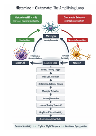
Picture 2: Hypothesis: overlapping factors that elevate histamine levels may act synergistically, thereby amplifying one another and contributing to a broad range of symptoms observed in ASD, ADHD, and PTSD. Illustration was generated using AI (ChatGPT 4.0.) and reviewed, corrected and completed by the author.
5.6 Glutamate–GABA Imbalance in ASD Pathophysiology
GABAergic and glutamatergic neurotransmission disruptions, which are symptomatic of glutamate–glutamine–GABA cycle failure, are regularly seen in biochemical investigations of ASD [333], [334]. GABA and glutamate activity and release are crucially modulated by redox signaling, which involves ROS and RNS. Post-mortem cortex tissue exhibits elevated glutamate, decreased GABA, and decreased kidney-type glutaminase activity [335]. Notably, ROS increase glutamate/GABA release and alter NMDA receptors, whereas redox pathways control GABAergic micro inhibitory postsynaptic current frequency, GABAA receptors, and GABA release [336]. According to [335], histological investigations reveal astrocytic structural abnormalities in ASD, suggesting reactive gliosis and poor neuroglial communication that worsen neurotransmitter dysregulation. In animal models of early-life mercury exposure, these neurochemical abnormalities, which are symptomatic of glutamate–glutamine–GABA cycle failure, are reliably reproduced. In these models, rats show increased extracellular glutamate, decreased GABAergic signaling, and down regulated glutamine synthetases. Importantly, by upsetting the delicate excitatory/inhibitory balance—a hallmark of ASD pathophysiology—Hg-induced astrocytic dysfunction, which is characterized by changes in astrocyte morphology and impaired neuroglial communication, critically underlies these observed ASD-like behavioural phenotypes [333], [337], [338], [339].
5.7 Histamine H3 Receptor and Acetylcholine
Acetylcholine (ACh) is released by neurons through an enzymatic reaction occurring at axon terminals and exerts broad neurotransmitter functions in the central, peripheral, and autonomic (sympathetic and parasympathetic) nervous systems, as well as at the neuromuscular junction, where it induces muscle contraction and thereby regulates the functional efficiency of many organs, such as the heart, lungs, intestines, urinary bladder, and endocrine and exocrine glands [340], [341]. Activation of histamine H3 receptors in the brain, for example in the hippocampus, may inhibit the release of the neurotransmitter acetylcholine (ACh) from cholinergic neurons, which are involved in cognitive processes and modulate attention, wakefulness, memory, and reward processing [342]. As a result, this may lead to impaired cognitive functions and memory in individuals with ASD. Low levels of acetylcholine have been investigated as a potential contributing factor to autism symptoms, as individuals with ASD exhibit certain abnormalities in the brain cholinergic system, including acetylcholine neurotransmission [343]. Enhancement of acetylcholine signaling in certain brain regions may improve cognitive functions, attention, and social interactions; however, excessive ACh activity in other areas may exacerbate sensory hypersensitivity and anxiety, worsen impulse control, executive functions, and emotional regulation [344]. There is also a feedback mechanism: acetylcholine, through its action on muscarinic or nicotinic receptors, can regulate mast cell function, leading to reduced histamine release under certain conditions (resulting in an anti-inflammatory effect of the cholinergic system) [345].
Many preclinical studies have demonstrated that histamine H3 receptor antagonists improve social functioning and reduce repetitive behaviors [203], [209], [270], [295], [297], [346], [347], [348], [349]. The antioxidant properties of H3 receptor antagonists clearly indicate that these compounds may represent a promising therapeutic option for the treatment of autism spectrum disorder (ASD) [350], [351], as well as other brain disorders in which microglia-driven neuroinflammation plays a central role, such as Alzheimer’s disease (AD), schizophrenia (SCH), and behavioral and psychological symptoms of dementia (BPSD)[200], ADHD, PTSD [352], [353].
6. Endothelium, Histamine and Thrombosis in Systemic Inflammation
The vascular endothelium plays a critical role in regulating histamine metabolism, coagulation, inflammation, and blood–brain barrier integrity through the balanced activity of histamine-synthesizing and histamine-degrading enzymes such as HDC, DAO, and HNMT [354]. When histamine degradation becomes impaired, excessive histamine signaling may promote endothelial dysfunction, platelet activation, coagulation cascade activation, vascular hyperpermeability, and immunothrombosis, potentially contributing to tissue hypoxia, neurological complications, stroke, myocardial infarction, and multisystem inflammatory responses [255], [355], [356], [357], [358], [359] also contributing to neuroinflammatory mechanisms through microglial activation, oxidative stress, and neurotransmitter dysregulation mediated by H1, H2, H3 and H4 receptors [129], [180], [181], [255]. Chronic inflammatory conditions such as obesity, diabetes, sepsis, and COVID-19 further amplify oxidative stress and endothelial injury, thereby increasing the risk of thrombotic [360] and neurovascular complications [361], [362], [11], [363], [364]. Moreover, ionizing radiation and oxidative stress may impair endothelial histamine-metabolizing capacity by reducing DAO and HNMT activity, leading to histamine accumulation, enhanced mast cell activation, blood–brain barrier disruption, cerebral edema, and intensified inflammatory and thrombotic responses [365], [366] often starting with DIC [367], which is associated with the sticking together of platelets and damage to erythrocytes that transport oxygen. Severe and prolonged hypoxia associated with impaired oxygen delivery in the context of thrombotic and inflammatory processes is associated with multi-organ dysfunction, including potential neurological impairments ranging from cognitive deficits to loss of consciousness, cardiac complications such as arrhythmias, heart failure, or myocardial ischemia [368], and renal and hepatic damage. In advanced or chronic cases, peripheral ischemic changes may occur, including cyanosis and tissue necrosis, which, under extreme conditions, can develop into gangrenous changes. In critically ill patients, such pathophysiological cascades can contribute to life-threatening conditions, especially when oxygen delivery remains inadequate due to persistent thrombosis [329] or systemic hypo-perfusion [369].
6.1 Importance of Methylation and Vitamins B6, B9, B12
Research shows that in both neurodevelopmental and neuropsychiatric disorders, including ASD, ADHD, PTSD, and related disorders, elevated histamine levels and impaired histamine degradation are associated with increased neural excitability, cognitive and emotional dysregulation, sleep disturbances, and multisystem symptoms [370], [371]. HIT and genetic variation in DAO and HNMT potentially exacerbate neuroinflammatory and behavioral symptoms [372], [373]. Histamine metabolism is mediated by DAO and HNMT. HNMT function plays a crucial role in neurodevelopment, as many mutations in the HNMT gene have been found in individuals with profound intellectual disabilities [374]. Methylation is a key biochemical process that involves the transfer of methyl groups (–CH₃) to molecules such as DNA (DMNT), neurotransmitters, and histamine, thereby regulating gene expression, detoxification, and neurotransmitter metabolism through SAM-dependent enzymes such as histamine N-methyltransferase (HNMT) [375], [164]. Therefore, proper methylation is essential for maintaining normal cellular and nervous system function. This system depends on one-carbon metabolism and adequate levels of vitamins B6, B12, and folate (B9) [810] to maintain SAM production [376], [377], [378], [379]. Importantly, a form of vitamin B6 called pyridoxal phosphate (PLP) plays a cofactor role for the enzyme glutamate decarboxylase (GAD), which consequently converts glutamate to GABA [380], [381]. PLP deficiency further reduces GAD activity, lowering GABA synthesis and potentially increasing glutamate, while simultaneously impairing HNMT function, which may lead to elevated histamine levels and neuroinflammation [380], [382]. Disturbances in this system may impair histamine and neurotransmitter degradation, increase homocysteine levels, and increase oxidative stress [383], thereby contributing to neuroinflammatory [384], [385], cardiovascular, and neurodevelopmental disorders observed in conditions such as ASD, ADHD, and other neuropsychiatric or neurodegenerative disorders [12], [26], [386], [387].
6.2 The Role of Neurotensin, CRH and Substance P in Inflammation
Neurotensin (NT) [388] and corticotropin-releasing hormone (CRH) are stress-related mediators that can activate mast cells [389] promoting histamine release and contributing to neuroinflammation and dysregulation of the gut-brain axis [390]. They link the nervous and immune systems, acting both centrally (HPA axis) and locally in tissues [391]. Both of these hormones have also been found to be elevated in conditions such as ASD [12], [26], [392], [81]. Similarly, substance P is a neuropeptide (a member of the tachykinin family) that activates mast cells, induces neurogenic inflammation, and promotes histamine degranulation, thereby linking stress, pain signaling, and inflammatory processes in the gastrointestinal and respiratory systems [393], [394], [81], [395]. Substance P has also been linked to several chronic diseases, such as obstructive pulmonary disease (COPD), asthma, anxiety, depression, nausea, and inflammatory bowel disease [396].
6.3 The Body’s Response to Antigens - Oral and Intravenous
To better understand these mechanisms, it is valuable to examine the pioneering lectures and discoveries of Nobel Prize–winning scientists who laid the foundations of modern immunology, neurophysiology, and histamine research, including Ivan Pavlov (neural digestive reflexes, 1904), Charles Richet (anaphylaxis, 1913), Sir Henry Dale (histamine and chemical neurotransmission, 1936), Daniel Bovet (antihistamines, 1957), and Sir James W. Black (histamine receptor pharmacology, 1988) (Nobel Prize lectures) [397].
6.3.1. Oral Intake and Mast Cell–Histamine Responses
The gastrointestinal tract represents the first stage of the body’s contact with nutrients delivered from the outside. It is a continuous tube running through the entire body, remaining in open communication with the external environment, which makes it, in a way, an “internally hidden” part of the body surface. Digestion occurs with the involvement of substances that retain their properties both in the digestive tract and outside the body under laboratory conditions, allowing them to be analyzed using chemical methods and described by the laws of chemistry. These substances are called enzymes, formerly known as ferments (Pavlov Lecture Nobel Prize) [398]. The process of digestion involving enzymes consists of the breakdown of food by enzymes secreted by the host organism in the gastrointestinal tract, enabling the absorption of nutrients. Fermentation, on the other hand, is the transformation of organic compounds (e.g., sugars) that occurs with the involvement of enzymes secreted by microorganisms (mainly bacteria and yeast), which break down organic substances (usually anaerobically), producing by-products such as organic acids or gases. Fermentation is used in many culinary processes, including sauerkraut production, yogurt and kefir preparation, and bread dough leavening, which promotes the formation of histamine (Fermentation) [399].
Histamine is one of the so-called biogenic amines—compounds formed from amino acids through the action of bacterial enzymes. Therefore, fermentation and histamine are closely related. During fermentation (e.g., pickling, cheese aging, wine fermentation), fermentative bacteria break down various compounds, including amino acids such as histidine, which can be converted into histamine through histidine decarboxylation by the bacterial enzyme histidine decarboxylase. Consequently, fermented products (aged cheeses, wine, beer, pickled vegetables, fish products) often contain elevated levels of histamine and other biogenic amines (tyramine, putrescine, cadaverine). In sensitive individuals (e.g., those with a deficiency of the DAO enzyme, which breaks down histamine), such products may trigger pseudoallergic symptoms (headache, urticaria, redness). A similar process occurs during putrefaction, where protein breakdown by bacteria leads to the release of biogenic amines. During spoilage of meat, fish, or cheese, bacteria produce histamine and other amines (putrescine, cadaverine), and consumption of histamine-rich food can cause histamine poisoning (e.g., scombroid poisoning from fish) or HIT.
Mast cells are present in human and animal salivary glands (parotid, submandibular, sublingual) as well as in the tonsils [400]. Their numbers increase, for example, during salivary gland inflammation, autoimmune diseases, or allergies. Upon contact with an allergen or food (e.g., milk, wheat, eggs, nuts, fish, strawberries, tomatoes, chocolate, seafood, aged cheeses, wine, fermented vegetables), mast cells undergo degranulation and release histamine and other mediators. The released histamine can modulate saliva secretion (acting on H1 and H2 receptors in the epithelium and blood vessels), participate in hypersensitivity reactions, and increase vascular permeability within the salivary glands, causing edema and inflammatory infiltration. Histamine plays a key role in the stomach, where enterochromaffin-like cells (ECL) secrete histamine, which binds to H2 receptors on the parietal cells of the gastric mucosa, stimulating the secretion of hydrochloric acid (HCl), the main component of gastric juice [401]; (Pavlov Lecture Nobel Prize) [398]. It has long been known that some people are sensitive to cheese, strawberries, fish, shellfish, eggs, raw meat, and even milk (lactose, casein A1). The symptoms observed in these individuals after consuming such foods are analogous to a histamine storm: acute abdominal pain, vomiting, diarrhea, colic, erythema, urticaria, severe itching, and sometimes heart problems, fever, and leukocytosis—these are anaphylactic phenomena. Anaphylaxis has become a common pathological phenomenon. “Anaphylaxis is the cause of adverse reactions that occur in a small number of people in response to certain proteins found in food.” (Nobel Prize Speed read: A shock response) [402]. Victor Hutinel, a Parisian pediatrician at the beginning of the 20th century, developed the term “food anaphylaxis,” which described analogous reactions and symptoms after ingestion of, for example, milk [403], [404], [405]. Wheat allergy associated with the risk of anaphylaxis represents a serious global food safety issue. Gliadins, glutenins, albumins, and globulins of wheat, as well as pesticides from spraying, can act as allergens and trigger severe allergic reactions in which histamine is released [406], [407], (Pavlov Lecture Nobel Prize) [398].
6.3.2. Potential Reason of Food Selectivity
These findings regarding food anaphylaxis may have significant clinical implications. Many cases of mild digestive discomfort, so-called indigestion, may result from mild anaphylactic reactions. It has long been believed that maintaining a regular, consistent diet is more beneficial than experimenting with diverse dietary patterns. Repeated consumption of the same protein may lead the body to gradually become accustomed to it, developing tolerance to this common antigen. This may explain why children with ASD often display so-called food selectivity, sometimes consuming only one or a very limited set of foods—possibly as a subconscious way to avoid triggering further histamine release. Analysis of numerous substances capable of inducing anaphylactic reactions, as well as those that can induce immunological tolerance, indicates that chemical and humoral diversity between individuals is practically limitless, highlighting the complexity and individuality of immune responses. It was previously assumed that organisms of the same age, race, and sex have identical body fluids. However, each organism possesses unique characteristics that make it distinct. Therefore, studying the physiology of the species alone is insufficient—understanding the physiology of the individual is necessary (Richet Lecture Nobel Prize) [399].
6.4 Intestinal Injection and Mast Cell–Histamine Responses
The human body is generally structured so that intact proteins are not expected to enter the bloodstream unless they are first broken down into smaller components by digestive enzymes (Nobel Prize, 1913) [408]. When foreign proteins do reach the circulation, the immune system reacts, triggering a defensive response that can lead to increased sensitivity upon subsequent exposure. It occurs when the immune system, after prior sensitization to an antigen such as food and food additives (e.g., monosodium glutamate, colorants) [406], venom [409], drugs (or various medications (e.g., depolarizing muscle relaxants, contrast agents, antibiotics, or latex) [410], or other substances, responds on re-exposure (repeated parenteral administration) [99], with IgE-mediated or idiopathic activation of mast cells and massive histamine release, it makes immune system more fragile and susceptible [411].
In 1906, Rosenau and Anderson demonstrated that anaphylaxis could be triggered not only by toxins but by many different proteins, even in extremely small amounts (0.00001 ml) given parenterally, through subcutaneous or intravenous injection in experimental animals, including milk, eggs, serum, plant or muscle extracts, dead microbial bodies, bacterial protein toxins, yeast cells (Richet Nobel Prize Lecture) [412]. The key observation was that introducing foreign proteins directly into tissues or the bloodstream could sensitize the organism and, upon re-exposure, trigger systemic hypersensitivity reactions (anaphylaxis) [413], (Richet Biography) [414]. Anaphylaxis may be triggered not only by classical allergens, but also by physical factors such as extreme temperatures (sometimes in combination with recently consumed food) [415], strenuous exercise, mechanical stimulation like vibration [80], [94], [416], and some vaccine adjuvants [417], [418], [419]. Symptoms range from mild itching and urticaria to severe systemic reactions such as hypotension, bronchospasm, edema, neurological impairment, and in severe cases to potentially life-threatening reaction, such as anaphylactic shock [420], also death [99]. Histamine is the key mediator of these processes, and treatment relies primarily on epinephrine, which counteracts its effects, with antihistamines used as supportive therapy [421].
There is a hypothesis that some components of intravenous vaccines entering and circulating in the bloodstream may activate mast cells in the endothelium and surrounding blood vessels, leading to excessive histamine release and additional activation of H1–H4 receptors, potentially contributing to chronic inflammation in multiple organs in the body. Some studies have suggested that, in susceptible individuals, immune activation following vaccination or exposure to certain antigens may contribute to excessive mast cell activation, histamine release, and chronic inflammatory or autoimmune-like symptoms. In a healthy individual, exposure to an antigen usually induces a balanced immune response, resulting in antibody formation and long-term immunological memory. In people in people with MCAS or with deficiency of HNMT or DAO enzymes [69], [70], [422], [423], exposure to vaccines components or other antigens may more easily trigger an exaggerated response, with strong mast cell activation and histamine buildup. This can result in allergic reactions, shock, or pseudo-anaphylaxis, where histamine plays a key driving role rather than a secondary one [69], [422], [424]. These significant differences in physiological responses are particularly important in the context of vaccination individuals.
These mechanisms are conceptually linked to classical research on anaphylaxis and hypersensitivity by Charles Richet and others, emphasizing that repeated exposure to specific antigens can, in predisposed individuals, trigger exaggerated immune responses mediated by mast cells and histamine rather than protective tolerance alone (Richet Nobel Prize Lecture) [412], [425], [426]. The concept of heightened reactivity to repeated exposure was described by Clemens von Pirquet in 1906, who introduced the term “allergy” to describe an altered immune response [427], [428]. Early research by Charles Richet and Paul Portier also helped establish the concept of anaphylaxis as a systemic hypersensitivity reaction to foreign proteins. Overall, these observations contributed to the understanding that the immune system can respond differently to repeated or systemic exposure to antigens, sometimes with severe reactions. Studies indicate that perivascular mast cells can capture IgE directly from the bloodstream through the vascular wall [429]. Bose et al. further demonstrated that cutaneous mast cells form cytoplasmic extensions reaching into the lumen of blood vessels [430], which may enable rapid mast-cell activation and intravascular degranulation after intravenous antigen or IgE exposure [431], [432]. The immune system may respond to a foreign protein or drug by producing antibodies that bind to the substance and form immune complexes. These complexes can accumulate in tissues, particularly within blood vessels, triggering inflammation, tissue injury, and the development of clinical symptoms. The reactions discussed above involve parenteral administration, where substances are delivered intravenously, subcutaneously, intrathecally, or intraperitoneally, bypassing the digestive tract. However, anaphylaxis may also develop after oral exposure, as food allergens absorbed through the gastrointestinal tract can trigger systemic immune and mast-cell activation (Richet Nobel Prize Lecture) [412]. In addition, histamine it participates in the development of SIRS and sepsis [11], which—like anaphylaxis—are systemic rather than local reactions [433]. In medical practice, vaccines are usually given in a monitored and controlled setting. In certain situations, preventive administration of antihistamines or other safety measures may be used to lower the likelihood of allergic responses or anaphylactic reactions.
6.4.1. Anaphylaxis
The phenomenon of anaphylaxis was first systematically described by Charles Richet and Paul Portier in 1902 titled On the Anaphylactic Action of Certain Toxins, and later classified as a form of immediate (Type I) hypersensitivity within broader immunological frameworks (Richet Nobel Prize lecture: Anaphylaxis, 1913) [412], [425], [426], [403]. Charles Richet received the Nobel Prize for his work on this phenomenon, inspiring further studies [434].
In most cases it involves prior sensitization with IgE antibodies against a specific antigen, although this is not obligatory, since reactions can also occur to individual components of a formulation. Clinically, it is classified into IgE-mediated anaphylaxis (occurring upon re-exposure), non–IgE-mediated pseudoanaphylactic reactions often triggered by excipients such as adjuvants, stabilizers or preservatives, and idiopathic anaphylaxis where no identifiable cause is found in a substantial proportion of patients [435].
This difference helps explain why the body responds differently to substances depending on whether they are ingested or introduced directly into the bloodstream, such as aluminum (Aluminium in blood) [436] or mercury. The same substance can have completely different effects depending on the route of administration: the intestines limit exposure, blood and tissues provide complete and rapid distribution, and the immune system responds more vigorously to systemic exposure.
Aluminium salts are commonly included in some vaccines as adjuvants, meaning substances that enhance and prolong the immune response to the antigen [437]. Aluminium is present in food, water, and some vaccines and is normally eliminated in small amounts via the kidneys, so it does not usually accumulate [438]. With higher or prolonged exposure, it may deposit in tissues and has been studied for potential neurotoxic effects and associations with neurological disorders, also such as Alzheimer’s and Parkinson’s disease [439], [128], [440], [441]; particularly in vulnerable populations [442], [443]. Some studies suggest a potential link between mercury exposure (as hapten bind with skin or plasma proteins [444], [445] and the presence of brain-reactive autoantibodies in individuals with ASD. It has also been reported that levels of these autoantibodies may correlate with both autism symptom severity and measured mercury concentrations in the blood [446], [447]. Reactions to aluminium and mercury [448] are generally non–IgE-mediated [449] and are more consistent with delayed inflammatory mechanisms rather than immediate allergic responses [450], [451], [452], often contributing to immune dysregulation and mercury-induced astrocytic dysfunction accompanied by autism-like behavioral characteristics [337], [339], [453]. Claims and statements of concern regarding vaccine components, including thimerosal, have been discussed publicly by various figures, including Robert F. Kennedy Jr. Jr., Secretary of the U.S. Department of Health and Human Services (HHS) [75], in recent speeches ((Mr. Kennedy speeches, 2025) [454], (Others speeches, 2025) [455]. Following extensive research and analysis, HHS adopted a recommendation to remove thimerosal from all U.S. influenza vaccines (HHS, July 23, 2025) [456]. Some examples from UK PIP Analysis data: E.R. Dalby, Carlos Alegria, "Analysis of UK New Claims for Personal Independence Pension (PIP) by Underlying Causes," data from UK DWP, ONS, and NHS vaccination statistics (UK PIP Results link) [815]. (Figure 1, 2,3,4,).
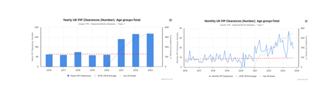
Figure 1: Hypersensitivity diseases. Yearly UK PIP Clarences. Cause nr: 176. Age group = Total.
Figure 2: Hypersensitivity diseases. Monthly UK PIP Clarences. Cause nr: 176. Age group = Total.
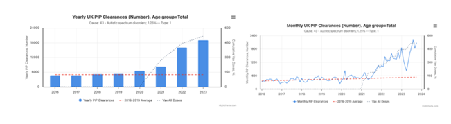
Figure 3: Autistic Spectrum Disorder 1,25%. Yearly UK PIP Clarences. Cause nr: 43. Age group = Total.
Figure 4: Autistic Spectrum Disorder 1,25%. Monthly UK PIP Clarences. Cause nr: 43. Age group = Total.
6.4.2. Intestines, DAO, Histamine, and ASD
A review article, “Histamine: A Mediator of Intestinal Disorders,” describes the role of histamine in gastrointestinal function, including its production, action through various receptors, and involvement in intestinal disorders, noting that in inflammatory states its levels are often elevated and may contribute to maintaining chronic low-grade inflammation [87]. In intestinal inflammation (e.g., food intolerances, Crohn’s disease, celiac disease, SIBO, intestinal infections) [457], the intestinal epithelium and enterocytes—the cells that produce the enzyme DAO (diamine oxidase)—are damaged [458]. In a healthy body, DAO neutralizes excess histamine in the intestine before it enters the bloodstream [459]. The level of diamine oxidase (DAO) measured in serum is an indicator of the integrity and condition of the small intestinal mucosa and may serve as a marker in diagnosing inflammatory bowel diseases, detecting allergic disorders, and liver cirrhosis (whose pathology is represented by changes in bacterial composition (gut dysbiosis) and SIBO (small intestinal bacterial overgrowth) [460], the risk of miscarriage, assessment of drug toxicity, and HIT [461]. Unfortunately, the cost of testing is high, and additionally, menstrual cycle, diet, medications, and sex may affect the reliability of the enzyme in diagnostics. DAO supplementation and a low-histamine diet can significantly help reduce symptoms of HIT and intestinal disorders [458], [462], [463].
Increased intestinal permeability and low-grade inflammation can secondarily reduce DAO levels [464]. The involvement of mast cells in regulating intestinal permeability and gut microbiota balance indicates their role in gastrointestinal disorders co-occurring with ASD [30]. These disorders also occur in older adults and in certain chronic diseases, which according to studies posed higher risk in COVID-19 infections [465] and higher mortality risk from COVID-19 in these three groups „ After the Elderly, Those with Intellectual Disabilities are at Greatest Risk of Death from COVID-19” [466], [467]. These studies support the author’s hypothesis that intestinal condition affects DAO levels, which in turn influence histamine levels, playing a significant role in the pathology of COVID-19, as well as in ASD, ADHD, and PTSD [10], [11], [468].
Changes in the gut microbiome and metabolites, such as propionic acid, can additionally disrupt the GABA–glutamate balance in the brain [469], exacerbating autistic behaviors [470]. Probiotic interventions show potential in modulating neurotransmitter balance and reducing excitation/inhibition imbalance in the nervous systems [471]. Studies suggest that disturbances in gut microbiota balance may play a role in the emergence of HIT symptoms. Further analyses are still needed to determine whether microbiota changes are a direct cause or rather a consequence of this intolerance [472] in [473]. In studies of individuals with ASD, the dominant symptoms of HIT were diffuse abdominal and head pain, recurrent diarrhea and vomiting, as well as skin rashes [474]. After starting a low-histamine diet (low in histamine-containing foods), symptoms resolved, and the use of H1 and H2 antihistamines was necessary only in two clinical situations [157]. Notably, in the human gastrointestinal tract, H1, H2, and minimally H4 receptors are present, whereas the H3 receptor is absent. It has also been observed that in patients with gastrointestinal diseases, the expression of these receptors changes [475], and under acute stress in rat gastrointestinal tracts, histamine content increases in colonic mast cells. This is associated with the release in the brain of interleukin-1 followed by corticotropin-releasing factor (CRF), because immune signaling between the gut and the central nervous system occurs via cytokines, and their levels depend on gut condition and microbiome composition [83]. Studies indicate that disrupted neuronal pathways in ASD may be modulated by mast cell mediators, such as histamine, serotonin, and cytokines, which influence both neuronal function and immune system responses, contributing to the development of characteristic symptoms of this disorder [476], [30]. In individuals with ASD and their mothers, autoimmunization is often observed, and mast cells can initiate and amplify these reactions by regulating lymphocyte numbers or releasing autoantigens [372].
6.5 Embryonic Development
New research directions also include analyses of prenatal environmental factors related to ASD occurrence, such as maternal use of antibiotics, analgesics, selective serotonin reuptake inhibitors, exposure to environmental toxins, immune, allergic, or metabolic disturbances, changes in gut microbiota, and IL-17 levels [477], [478], elevated levels of hormones, biogenic amines (including histamine), and oxidative stress – all influencing the development of the child’s brain and the later emergence of ASD. These potential biological mechanisms may serve as preventive strategies [479].
6.5.1. Mother Inflammation During Pregnancy
During pregnancy, inflammatory processes in the mother disrupt the normal function of the placenta, increasing the likelihood of neurodevelopmental disorders in the child [480]. In mice, induced maternal inflammation leads to increased conversion of tryptophan to serotonin in the placenta, which disrupts the development of serotonergic neurons in the fetal brain; as a result, the fetal forebrain is exposed to elevated levels of this biogenic amine, leading to characteristic changes in key neurogenic processes regulated by serotonin (5-HT) [481], indicating a mechanism by which maternal inflammation can affect brain development in the child [482]. Furthermore, in a decade-long cohort study involving 555 mother–child pairs, the presence of specific proinflammatory proteins during pregnancy correlated with an increased risk of neurodevelopmental disorders in offspring [483]. Other studies confirmed that mothers with asthma, diabetes, or extreme obesity during pregnancy had a very high likelihood of giving birth to a child later diagnosed with ASD [484]. Another study of 1,241 mother–child pairs showed that elevated levels of selected inflammatory cytokines in the placenta and umbilical cord blood were associated with developmental delays, and that the negative impact of a proinflammatory intrauterine environment on children’s neurological development was exacerbated by low maternal vitamin D levels [485]. Another studies shows that supplementation of vitamin D has effects on interleukin -6, serum serotonin and on reduction of core symptoms in children with ASD [486], [487], better results were dependent of the earliest age of this treatment [488].
6.5.2. Histamine and Embryonic Brain Development
Histamine appears very early in the developing brain and influences neural stem/progenitor cells depending on the type of receptor activated. Scientific studies provide a broad overview of the role of histamine in brain embryogenesis [489]. These studies present the dynamics of histamine levels during the prenatal period, its effects on developing brain cells, and potential links to developmental disorders [200], [490]. A classic study describing HNMT activity in the pineal gland, thalamus, and peripheral nerves during chick development shows the appearance of enzyme activity already in the embryo and its increase with maturation. It covers the development of enzymatic activity that methylates and degrades histamine [491]. Research shows that measurements of DNA methylation during pregnancy, as an important epigenetic feature, may correlate with the later development of ASD in children [492].
A better understanding of epigenetic mechanisms also allows comprehension of how the developing epigenome is susceptible to genetic and environmental influences (for example, factors that raise histamine levels) [493], [494], including already in the placenta [495]. For example, altered GAD1 methylation patterns in ASD may affect its expression and reflect heterogeneity in epigenetic regulation at the cellular level [496], while higher methylation of the oxytocin receptor OXTR in intron 1 observed in adults with ASD compared to neurotypical individuals has generated great interest in social neuroscience and psychiatry regarding oxytocin’s role in social cognition, and OXTR hypermethylation as a biomarker for adults with ASD [497].
For DNA/histone methylation, the main methyl group donor in the body, SAM (S-adenosylmethionine), is required – which also supports histamine breakdown via HNMT. Proper levels of, among others, folic acid, vitamin B6, and B12 support the synthesis of S-adenosylmethionine (SAM) – a key methyl group donor in the CNS – thereby exerting a neuroprotective effect by limiting oxidative stress, histamine levels, excessive microglial activation, and neuroinflammation. In the case of their deficiency, SAM levels decrease while SAH (S-adenosylhomocysteine, which acts as an inhibitor of methyltransferases) increases, leading to impaired DNA and protein methylation, slower histamine degradation (particularly via HNMT), and consequently elevated histamine levels in tissues and body fluids. This may result in symptoms of HIT, allergic hyperreactivity, migraines, and may also contribute to the pathogenesis of certain neuropsychiatric disorders (ASD, depression) [498]. High histamine levels may signal a burden on the DNA methylation cycle, thereby influencing the epigenetic regulation of genes in the embryo. Additionally, signs of oxidative stress and disturbances in DNA methylation processes are frequently observed in individuals with ASD [498]. Supplementary therapy studies indicate that supplementation with components such as vitamins B6, B9, B12, C, E, D, omega‑3 fatty acids, glutathione, and choline can effectively influence methylation regulation and nutrition, support metabolic functions, and significantly alleviate ASD symptoms [499].
7. Histamine Metabolism – the Enzymes HNMT and DAO
The enzymes DAO and HNMT are crucial for histamine degradation. DAO functions extracellularly, breaking down histamine derived from the diet. HNMT works intracellularly and is the main enzyme responsible for histamine breakdown in the central nervous system. HNMT methylates histamine (N‑methylation), whereas DAO oxidatively deaminates it, producing different metabolites. Reduced activity of these enzymes can result in excess histamine and symptoms that exacerbate autistic behaviors in ASD [168], [164].
7.1 HNMT
HNMT (histamine N-methyltransferase) is an enzyme responsible for the intracellular degradation of histamine. The highest HNMT activity is observed in the central nervous system —particularly in the brain—as well as in the liver, kidneys, and lungs. HNMT is a monomer with a molecular weight of approximately 33 kDa and is dependent on the presence of magnesium ions (Mg²⁺) and SAM (S-adenosylmethionine) as a methyl group donor. HNMT catalyzes the methylation of histamine, leading to the formation of N-methylhistamine. This is the main pathway of histamine degradation in the CNS, where DAO is not present. HNMT regulates histamine levels in the brain and thus influences processes related to sleep, memory, learning, and emotional responses [373], [93]. Polymorphisms (genetic variants) in the HNMT gene may lead to altered enzyme activity and are being investigated in the context of neurological and psychiatric disorders such as ADHD, Parkinson’s disease, and schizophrenia [500], as well as autism [39]. Research results indicate that HNMT plays an important role in human neurological development, and its deficiency may increase sensitivity to histamine (HIT), particularly in brain tissues. Reports of mutations in the HNMT gene in humans and their associations with intellectual developmental disorders provide evidence that the HNMT gene has functional significance in the development of the nervous system [501].
7.2 DAO
DAO is an enzyme belonging to the oxidase group; it is an enzymatic protein composed of an amino acid chain—therefore, it is a globular protein. DAO is produced mainly by the epithelial cells of the small intestine (enterocytes), but it is also present in the kidneys, liver, placenta, and to a lesser extent in other tissues, where it breaks down and neutralizes histamine. DAO is a dimer, and its activity depends on proper expression of the AOC1 gene and the availability of cofactors. Its structure contains an active site with a copper-containing coenzyme, which participates in the oxidation reaction [452], [453]. The DAO enzyme plays a key role in catalyzing the breakdown of histamine and other biogenic amines (e.g., putrescine, cadaverine) [454], converting them into the corresponding aldehydes, ammonia, and hydrogen peroxide [455]. DAO catalyzes the oxidative deamination of histamine, leading to the formation of imidazole acetaldehyde, which is then further metabolized [452]. Its main function is the degradation of histamine derived from the diet (exogenous histamine, e.g., from fish, cheeses, wine, and fermented foods), thereby protecting the body against excess histamine supplied with food [455].
The DAO enzyme may also be released by heparin [456]. The body’s response to heparin in the form of increased DAO levels is poorly predictable and strongly depends on individual characteristics [457]. DAO deficiency or low DAO activity may lead to the accumulation of histamine and symptoms of HIT, such as headaches, diarrhea, urticaria, skin flushing, and even allergy-like reactions, so-called pseudoallergies [11], [458]. DAO activity may decrease in inflammatory bowel diseases (e.g., celiac disease, Crohn’s disease, SIBO), with damage to the intestinal mucosa, under oxidative stress, due to certain medications or toxins that inhibit DAO, when DAO is continuously utilized in the presence of excess histamine, or when cofactors (copper, vitamin B6, zinc) are deficient. In practice, this means that chronically high histamine levels may “overload” DAO, causing its efficiency to decrease and histamine to accumulate in the body [372].
Serum DAO activity differs significantly by sex, with women showing higher serum DAO levels than men, what is shown in article “Sex is the main factor determining DAO activity in healthy individuals” [509]. Some studies have shown that individuals with ASD may have lower activity of the DAO enzyme, which can result in excess histamine and symptoms that exacerbate autistic behaviors (e.g., anxiety, aggression, sleep disturbances) [168]. Low blood DAO levels may indicate a reduced capacity of the body to degrade histamine—for example, in HIT or in cases of damage to the intestine, liver, or kidneys. High blood DAO levels are sometimes observed during pregnancy (the placenta produces DAO) or as a result of enzyme release from tissues in certain disease states.
Histamine induces estrogen production in the ovaries, while estrogen induces the release of histamine from mast cells in the uterus [85]. For example, women with endometriosis have high histamine levels and a tendency toward allergies, which suggests reduced DAO levels [184]. When there is an excess of histamine in a woman’s body, it may increase estrogen levels and disrupt hormonal balance. Histamine additionally intensifies uterine contractions and activates pain receptors [510], [511], thereby increasing sensitivity to menstrual pain, cyclic headaches, and digestive problems [183]. However, not all women who experience these symptoms suffer from HIT. Before menstruation, the body demands an increased amount of histamine in order to intensify uterine contractions, which during menstruation are a natural mechanism for clearing the uterus of unnecessary tissue and enabling the start of a new menstrual cycle (PMDD and PMS) [512], [513], [514], (IAPMD) [515]. Therefore, before their period, women experience cravings for histamine-rich foods such as chocolate, cheeses, ketchup, and herring, and the physiologically required high level of histamine at that time causes irritability, nervousness, mood swings, headaches, sleep problems, bloating, and abdominal pain—so-called PMS (Premenstrual Syndrome) [516], [517], [518]. Serum DAO levels vary depending on the menstrual cycle; studies indicate that plasma DAO concentrations are significantly lower in the follicular phase than in the luteal phase [519]. In addition, DAO produced in the placenta plays an important role in female reproductive functions, including the maintenance of pregnancy [184].
7.2.1. During Pregnancy, the Placenta Produces 500 Times More DAO
A review of the role of histamine in embryo–uterus interactions, proliferation, and differentiation during pregnancy indicates that both excessively low and excessively high histamine levels may affect the course and development of pregnancy. Prenatal exposure to stressors (such as infections or toxins) may simultaneously influence the development of the embryonic immune and nervous systems [490], including stimulation and sensitization of mast cells and the development of MCAS (i.e., frequent or continuous histamine release) [459]. In addition, estrogens affect mast cell function (via estrogen receptors) and may increase their tendency to degranulate, thereby increasing local histamine concentrations. This explains observations of “estrogen → increased histamine” in the context of allergies, PMS, or pregnancy [514].
Scientific studies report that during pregnancy the placenta produces large amounts of the enzyme DAO (diamine oxidase); it is estimated that the placenta may produce up to 500 times more DAO than other tissues, such as the intestines or kidneys, depending on the stage of pregnancy and individual differences [184], [520]. Within the first 20 weeks of pregnancy, DAO levels measured in maternal plasma increase exponentially—up to 1000-fold compared with pre-pregnancy levels [521]. This high production is crucial for histamine metabolism in both the mother and the fetus, protecting against excessive histamine levels that could harm the developing organism, specially with Rh or ABO blood group incompatibility [185]. DAO in the placenta degrades histamine derived from the maternal diet or released during immune reactions, preventing its transfer into the fetal circulation. This is particularly important because the fetus has an immature histamine-metabolizing system. It has also been observed that while DAO activity in the mother’s body shows a steady upward trend, only minor fluctuations in its concentration occur in amniotic fluid and fetal blood [522]. Histamine concentrations in maternal blood during normal pregnancy fall below levels observed in healthy non-pregnant women [185]. In contrast, in complicated pregnancies, a halt in the increase of maternal plasma DAO levels and an elevation of circulating histamine levels are observed, significantly increasing the risk of preeclampsia, threatened miscarriage, and spontaneous miscarriage [523], [524].
High expression of the AOC1 gene in the placenta ensures increased DAO enzyme activity, which represents an adaptation to physiological changes in pregnancy such as increased vascular permeability and inflammatory responses. Certain genetic mutations, such as polymorphisms in the AOC1 gene (encoding DAO) or the HNMT gene, may lead to deficiencies of these enzymes and elevated histamine levels [39]. These genetic variants have also been observed in some children with autism spectrum disorder (ASD) [154]. DAO deficiency in a pregnant woman combined with exposure to environmental stressors—external stimuli, trauma, or histamine-rich or histamine-releasing foods—may increase the risk of pregnancy complications and potentially affect child development [525]. Maintaining a balance between histamine and DAO plays a key role in the normal course of pregnancy and the health of the child [184]. When most histamine in the mother’s body has been degraded by DAO, pregnant women may intuitively reach for so-called “cravings,” i.e., histamine-rich foods such as herring, chocolate, or pickled cucumbers, in order to improve digestion.
Immediately after childbirth, when the placenta is removed, DAO levels drop sharply and return to pre-pregnancy values. This means that the body loses the additional “buffer” protecting against excess histamine. As a result, histamine may act more strongly for some time, which in some women manifests as increased sensitivity to histamine-rich foods or histamine-releasing stressors. Symptoms attributed to histamine sensitivity after childbirth may include headaches, migraines, fatigue, skin problems (itching, urticaria), hair loss, and mood disturbances (postpartum depression may be partially modulated by neurotransmitters and histamine, although this is a complex mechanism) [526], [527], [184].
7.2.2. DAO, Cooper and A2 Casein in Breast Milk
The enzyme DAO (diamine oxidase) is present in human breast milk, although its concentration is relatively low compared to other tissues, such as the placenta, which produces it in much larger amounts. The presence of DAO in breast milk is important for histamine metabolism in infants, particularly during the first months of life, when their own enzymatic system is not yet fully developed (Centers for Disease Control and Prevention) [528] and may have a protective effect [529]. DAO activity is highest in colostrum, and significant DAO activity is also observed in mature human milk (up to 30 days of lactation), which may be related to the need for histamine degradation during early lactation [530], [531]. The highest concentrations are generally observed around the peripartum period [529]. Research indicates that DAO in breast milk also originates from the secretions of mammary glands [529] and may support the breakdown of histamine provided by the mother’s diet or produced endogenously by the infant [157]. In newborns and infants, the digestive system—including DAO production—is immature, making the presence of this enzyme in breast milk crucial for protection against excess histamine, which could trigger allergic reactions or inflammation [532], [533]. However, in cases of mastitis, higher concentrations of histamine, spermine, and putrescine are found in breast milk [534], which may lead to HIT symptoms in children [165], [535]. DAO levels in milk are variable and depend on the stage of lactation, the mother’s diet, and her individual expression of the AOC1 gene (which encodes DAO). DAO concentrations in milk are insufficient to fully compensate for enzyme deficiency in children carrying AOC1 gene variants (e.g., children with HIT). In such cases, the mother’s diet (avoiding histamine-rich foods) may be crucial [527]. It should be noted that "in lactating rats, all newly absorbed copper is preferentially directed to the mammary gland, where it is delivered at higher concentrations to the milk" [530]. Copper-transporting ATPases are expressed on mammary epithelial cells, but according to the researchers, "their role in delivering copper to the milk has not been clarified" [536]. According to the author's hypothesis, if we know that copper is a cofactor for the enzyme DAO, and the baby is no longer protected by this enzyme from the placenta after birth, then perhaps it must be supplied in large quantities so that the baby's body can neutralize histamine, which supports the statement that "in mammals, adequate copper supply during early development is critical, because inadequate copper during pregnancy and early infancy can be fatal" [536].
After weaning, the child’s DAO levels begin to depend on their own production, which gradually develops over the first years of life [165]. A 2006 study found that children who were not breastfed were at significantly increased risk of developing autistic disorders. The same applied to children fed formula without simultaneous supplementation with docosahexaenoic acid (DHA) and arachidonic acid (AA). The results were compared to children who were breastfed for more than six months, suggesting that longer breastfeeding or supplementation with DHA/AA is associated with a reduced likelihood of ASD [537]. These findings align with information that human milk (breast milk) contains exclusively A2-type casein, as do goat, sheep, buffalo, and camel milk—proteins that are more natural for humans and better tolerated by the digestive system [538].
7.2.3. Lactose, A1 and A2 Casein in Milk
Lactose is the so-called milk sugar—a natural disaccharide found in milk and dairy products. In order for lactose to be absorbed, the body requires the enzyme lactase (β-galactosidase), which is produced in the small intestine. Lactase hydrolyzes lactose into its constituent monosaccharides, glucose and galactose, allowing their subsequent absorption through the intestinal wall into the bloodstream. When lactase is absent or insufficient, lactose is not digested and passes into the large intestine, where it undergoes bacterial fermentation, which can increase intestinal permeability, indirectly activate mast cells, and exacerbate histamine-related symptoms. Accompanying symptoms include bloating, abdominal pain, cramps, diarrhea, nausea, and a feeling of “sloshing” in the stomach. This is not an allergy, but an enzymatic lactose intolerance [539]. Casein is a group of milk proteins to which the human body can respond differently (inflammatory or allergic reactions). In humans, casein constitutes about 20–40% of milk proteins, with the remainder being whey proteins. In cow’s milk, casein makes up about 80% of the protein content [540]. Casein is highly valuable because it contains phosphorus, calcium, and proteins, forming a casein clot in the stomach that is digested slowly over many hours, providing a steady stream of amino acids, which are used, among other things, in hemoglobin synthesis (the oxygen-carrying protein) [541]. It has been observed that some people who are allergic or sensitive to various cow milk casein proteins tolerate goat milk well. One explanation may be the A1/A2 hypothesis, referring to variants of beta-casein that differ by only a single, but significant, amino acid at position 67 [540].
A2 casein (with proline at position 67) occurs naturally in almost all mammals. However, in European dairy cattle, a genetic mutation occurred (A1/A1 or A1/A2), which changed the protein structure to A1 casein (with histidine at position 67) [542]. The A1/A1 or A1/A2 mutation, due to higher milk yield, was propagated through selective breeding, and only a few cows carry the A2/A2 genotype, producing milk containing only A2 casein. Cows with the A2/A2 genotype are most commonly found in developing countries, mainly in Asia and Africa. Keith Woodford, in his book “Devil in the Milk”, compiled over 100 scientific publications linking A1 casein with increased risks of various health issues, including skin problems, joint pain and inflammation, diabetes, heart disease, autoimmune disorders, mental health issues, autism spectrum changes, and schizophrenia [543]. For this reason, consumption of milk containing A2 casein is recommended (also demonstrated in mouse studies) [544], [545]. This small difference in the amino acid of the A1 and A2 β-casein molecule is highly significant [546], because A1 and A2 β-casein interact differently with digestive enzymes in our stomach and intestines [547].
During digestion of A1 casein (with histidine), the stomach produces the peptide beta-casomorphin-7 (BCM7), which has pro-inflammatory and opioid-like effects (similar to morphine or heroin), meaning it acts as a drug and toxin [548]. This peptide can negatively affect the digestive, nervous (brain function), hormonal, and immune systems, potentially exacerbating autism symptoms [549]. These facts and observations support the author’s hypothesis regarding the important role of histamine in ASD pathophysiology. They suggest that not only the BCM7 peptide (as a biologically active toxin) can induce histamine and cytokine release, leading to gut inflammation, which promotes further histamine degranulation from mast cells and impairs histamine breakdown by DAO (diamine oxidase), but also that histidine in A1 casein can, through decarboxylation in the gastrointestinal tract by bacterial or tissue enzymes, be converted into histamine. Excess histamine may slow intestinal motility, increase intestinal barrier permeability, and exacerbate symptoms observed in various medical conditions [550]. A1 casein milk can indirectly increase histamine levels in the body. As a result, sensitive individuals (e.g., with HIT, gut disorders, or reduced DAO activity) may experience symptoms similar to histamine excess or HIT after consuming it [547]. (Table 2).
Table 2: Secondary reactions in the intestine and tissues – elevated histamine levels and intensified histamine response.
|
Phenomenon |
Mechanism |
Final effect |
|
Inflammation |
BCM-7 activates opioid receptors and NF-κB |
Cytokine and histamine release |
|
Mast cell activation |
Mast cells respond to inflammatory stress |
Histamine released locally and systemically |
|
Intestinal barrier damage |
Increased intestinal permeability |
Endotoxins and allergens penetrate into the bloodstream, intensifying the histamine response |
|
Decrease in DAO activity |
DAO (diamine oxidase) breaks down histamine, but BCM-7 and intestinal stress inhibit it |
Histamine is not broken down – its level rises |
|
Histidine in A1 casein |
Histidine decarboxylase, the gut microbiota is disrupted, and the intestinal environment promotes fermentation and decarboxylation |
Converted into additional histamine |
Summary of the possible mechanism:
A1 milk (β-casein with histidine) → Digestion in the gut → Formation of BCM-7 → Increased intestinal permeability ↑ + inflammation ↑ → Mast cell activation ↑ → Histamine release ↑ → DAO inhibition ↓ → Histamine is not broken down → Rise in body histamine levels (intolerance symptoms) ↑ → Possible histamine excess symptoms from A1 milk → headaches, migraines, runny nose, shortness of breath, nasal congestion, rapid heartbeat, skin itching, hives, bloating, diarrhea, reflux, anxiety, insomnia ↑ → Inflammation ↑ → Impaired development ↓
Studies have shown that children fed milk containing A1 casein also develop more slowly than those fed milk with A2 casein [551], and A1 casein has also been linked to SIDS (sudden infant death syndrome) and other health issues in infants [552]. Therefore, A2 human milk is considered safe for infants, including those sensitive to A1-type cow milk proteins [553], [554].
7.2.4. Neonatal Sepsis and Hyperbilirubinemia (Jaundice) in Newborns
Researchers suggest that neonatal sepsis is primarily related to immunological immaturity, particularly in premature infants and boys [555], supporting the hypothesis proposed by Filcek [11]. Although sepsis is suspected in 7–13% of newborns, only 3–8% of cases demonstrate confirmed bacterial infection [556], suggesting that many newborns may experience sepsis-like inflammatory responses associated with excessive histamine signaling, DAO reduction (placenta removal) and antibiotic-induced [556] (damaging the intestines where DAO is produced), and excessive mast cell activation caused by invasive hospital procedures commonly used in premature infants [556], [557]. The author hypothesizes that loss of placental DAO after cord clamping combined with labor (prolonged compression activates mast cells in the skin), a change in ambient temperature from 37 °C (inside belly) to 18°C (outside), cold air in the lungs, and hospital environmental factors may induce mast cell degranulation, resulting in increased histamine release, with time leading to histamine storm, activating also histamine receptors H1-H4. This cascade can contribute to microthrombus formation, erythrocyte hemolysis, hyperbilirubinemia, jaundice, impaired circulatory function, and, in some cases, neurological complications, multi-organ inflammations, symptoms that resemble sepsis [11], [558], [559], [560], [561], [562].
Clinicians should consider important factors that could increasing the risk of neonatal sepsis [563] and implement appropriate preventive measures [564]. Prolonged recovery from sepsis may be associated with the risk of neurodevelopmental disorders [564], as well as with elevated bilirubin levels [565], the excess of which in severe neonatal jaundice [563] can lead to brain dysfunction [566]. It has also been shown that elevated unconjugated bilirubin levels has neurotoxic effects and may contribute negatively to brain tissue also cause the neurological damage [567], [568]. The author hypothesizes that erythrocyte aggregation [569] may be associated with microthrombosis and disseminated intravascular coagulation (DIC), potentially induced by excessive histamine levels and secondary hematological abnormalities, including thrombocytosis. These mechanisms may lead to increased erythrocyte breakdown and increased bilirubin levels, manifesting as jaundice of the skin and sclera, similar to those observed in sepsis [11], [570], [571]. This mechanism may be further enhanced by body’s estrogens, estrogens from environmental, and other toxic factors that activate mast cells to release histamine [559], [181], [560], [561], [514]. The hypothesis is that loss of placental DAO after birth, in combination with environmental factors and potential blood group incompatibility, may lead to mast cell degranulation, increased histamine levels, congenital metabolic disorders, microthrombi formation, erythrocyte disintegration, hyperbilirubinemia, jaundice (which appear after 2-3 days of life), exacerbate the inflammatory response through histamine release and activation of H1–H4 receptors [555] and potentially SIRS, sepsis or brain damage [569].
Biological, chemical, physical, environmental factors → mast cell degranulation → histamine release → microthrombus formation → erythrocyte hemolysis → hyperbilirubinemia → jaundice → impaired circulatory function, neurological complications, multiorgan inflammations → symptoms that resemble SIRS and sepsis.
8. Histamine Pathway Genes and Environmental Factors Play a Role in ASD
The histaminergic nervous system (HS) is responsible, among other things, for regulating cognitive functions and behavior, and it is associated with various neurological disorders such as Tourette syndrome (TS), which is frequently observed in individuals with ASD [572]. This suggests a significant role for histamine in the development of ASD symptoms [259]. The hypothesis presented suggests that identifying and understanding the physiological role of disturbances in the histaminergic system (HS) could indicate potential directions and key insights for the development of prevention strategies and new therapeutic approaches in ASD, which may also involve both genetic and environmental factors [150]. Interactions between genetic and environmental factors influence the immune system, leading to cytokine imbalance, which, as research shows, occurs in ASD and, together with MCAS and histamine, may underlie ASD [573]. This is particularly relevant because inappropriate or excessive immune activity, including cytokines activated by histamine receptors, can significantly affect the nervous system and have neurological consequences [265].
Key genes that influence histamine regulation in the body include: HDC, which affects endogenous histamine production; genes involved in histamine and metabolite breakdown, including AOC1 (encoding DAO), HNMT (encoding HNMT), MTHFR (C677T and A1298C mutations), which helps regulate methylation necessary for reducing intracellular histamine via HNMT; PEMT, which indirectly supports mast cell stabilization; as well as CYP2C19 (implicated in histamine metabolism Diamine oxidase (histaminase)), COMT (Important for catecholamine and phenol metabolism [574], MAOB, CYP3A4 (important for the metabolism of most drugs), PST (Phenol sulfur transferase - important for metabolism polyphenols) and ALDH7A1 [575], [576]. Gene Ontology classification lists over 200 genes participating in the histamine pathway, encoding other enzymes and proteins [577]. Polymorphisms in genes encoding histamine-degrading enzymes influence individual differences in histamine metabolism and may predispose to various diseases [578], [579]. For example, genetic variants in AOC1 and HNMT affect autism spectrum disorders [39], as well as Parkinson’s disease and schizophrenia [500]. These genetic variations, together with environmental factors that activate the immune system, support the histaminergic hypothesis in the development of neurodevelopmental disorders [580], [581] as well as in systemic diseases [582].
8.1 AOC1 Gene of the DAO Enzyme
The AOC1 gene is located on chromosome 7q36.1 in humans. It encodes the DAO protein, which is a copper-containing enzyme belonging to the amine oxidase family. DAO catalyzes the oxidation of biogenic amines such as histamine, putrescine, and cadaverine, converting them into less active metabolites, for example imidazole-4-acetic acid from histamine. AOC1 expression can be modulated by dietary factors, inflammatory states, or genetic polymorphisms. Mutations or variants in AOC1, for example rs10156191 and rs1049742, are associated with reduced DAO activity, which may lead to HIT [154]. Studies suggest a link between AOC1 polymorphisms and ASD or autoimmune diseases, where DAO deficiency may exacerbate inflammation and allergies [583], [578]. DAO deficiency causes histamine accumulation, which manifests as migraines [584], urticaria, sleep and digestive problems [88].
8.2 HNMT Gene of the HNMT Enzyme
The HNMT enzyme (histamine N-methyltransferase) is encoded by the HNMT gene, located on chromosome 2q22.1 in humans. It is the main enzyme responsible for the intracellular metabolism of histamine in the central nervous system and the brain [581]. HNMT catalyzes the methylation of histamine, converting it into the inactive metabolite N-methylhistamine with the help of the cofactor S-adenosylmethionine (SAM) [167]. HNMT expression is regulated at the transcriptional level and can be modulated by factors such as inflammatory states, drugs (e.g., methylation inhibitors), or genetic polymorphisms. Genetic variants, such as rs11558538 (Thr105Ile), are associated with reduced HNMT enzyme activity [585], [586]. Polymorphisms in HNMT are being studied in the context of allergies, asthma, migraines, autism spectrum disorder (ASD), and schizophrenia, where impaired histamine methylation may exacerbate inflammation or neurotransmission [587], [501], [588]. Genetic deficiencies such as polymorphisms in the AOC1 and HNMT genes [587] as well as NOS2, or functional deficiencies acquired, for example through intestinal inflammation, lead to histamine accumulation, exacerbating neuropsychiatric symptoms [154].
8.3 NOS2 – Gene for iNOS
Studies suggest that insufficient expression of AOC1 and NOS2 determines susceptibility to disease, increasing the risk of infections, and also participates in the development of COVID-19 [589]. Additionally, it is important to note the NOS2 gene, which encodes inducible nitric oxide synthase (iNOS), located on chromosome 17q11.2-q12 [590]. The iNOS enzyme is responsible for producing large amounts of nitric oxide (NO) in response to infection, stress, inflammatory, and immunological factors. It plays a key role in defense mechanisms, inflammatory processes, and sepsis, but excessive NO production can also lead to tissue damage. NOS2 polymorphisms (e.g., rs2297518) may affect NO regulation, which is associated with the risk of asthma, ulcerative colitis, and major common diseases in intensive care units, including the development of SIRS and sepsis [591], acute lung injury, and multiple organ failure [592]. NO is also being studied in relation to susceptibility to cardiovascular disease, diabetes, cancer, and neuroinflammatory diseases [593]. High iNOS expression is often associated with excessive NO production, leading to nitrosative stress and subsequent mitochondrial and neuronal damage [594]. Hyperactive NOS2 stimulates iNOS to continuously produce large amounts of NO, independently of calcium concentration [592]. Under oxidative stress, this NO reacts with free radicals, especially superoxide anion (O₂•⁻), forming peroxynitrite (ONOO⁻), a highly reactive and toxic oxidant capable of damaging lipids, modifying proteins, and inducing DNA damage [595]. This mechanism is highly relevant in sepsis, neurodegenerative diseases such as Parkinson's and Alzheimer's [596], autoimmune diseases, and chronic inflammatory conditions [593]. Research on peroxynitrite decomposition offers hope for restoring cardiovascular function in sepsis [591].
8.3.1. Common Biological Pathways: Histamine and NO, and NMDA Receptors
Histamine, through H1 and H2 receptors, activates endothelial nitric oxide (NO) production [597], [598]. As a result, NO acts as a vasodilator, which explains why histamine causes decreased blood pressure, skin flushing, and increased blood flow during inflammation and allergic reactions [599]. When histamine is not properly degraded, for example due to HNMT or DAO polymorphisms, it enhances inflammatory responses, which can sustain NOS2 expression. Both histamine (in cases of HNMT deficiency) and NO (in cases of NOS2 overexpression) contribute to neuroinflammation, neurotoxicity, and impairments in memory, concentration, and psychiatric symptoms such as ASD, PTSD, and depression [596], [93]. A shared mechanism is glutamate excitotoxicity: histamine and NO modulate the activity of NMDA receptors (N-Methyl-D-Aspartate receptors), which are one of the main receptor types for glutamate, the primary excitatory neurotransmitter in the brain [600]. Excessive NMDA activation leads to overproduction of NO, causing nitrosative stress and neurotoxicity. Additionally, NO acts retrogradely on presynaptic terminals, increasing glutamate release and further amplifying excitotoxicity. NMDA modulation is observed in Alzheimer’s disease [601] and schizophrenia [602]. NMDA antagonists or agonists are used therapeutically [593].
Inflammatory states and mast cell activators → released histamine → stimulates endothelium and immune cells → increased NOS2 expression → iNOS induction in response to inflammation, infection, or immune stress → more NO → excessive NMDA activation → overproduction of NO + glutamate release → excitotoxicity → neuroinflammation in the brain → neurodevelopmental and neurodegenerative diseases.
8.4 Genetic Associations and Clinical Significance
The genes AOC1, HNMT, and NOS2 are functionally connected through a shared pathway: mast cells → histamine → NO.
- Polymorphisms in AOC1 (e.g., rs10156191, rs1049742) are associated with reduced DAO activity → higher histamine levels in the body and brain → histamine intolerance
- Polymorphisms in HNMT (e.g., rs11558538 – Thr105Ile) → lower enzyme activity → higher histamine levels in the brain.
- Polymorphisms in NOS2 (e.g., rs2297518) → iNOS overexpression → increased NO → higher glutamate levels in the brain [811].
When disruptions occur simultaneously in these genes → a synergistic effect arises: chronic inflammation, oxidative stress, neuronal damage, and neurotoxicity [68], which has clinical relevance in:
ASD, ADHD, and PTSD: Studies indicate that both excessive histaminergic activity (HNMT ↓, DAO ↓) and NO overproduction (NOS2 ↑) contribute to deficits in attention, memory, and emotional regulation [37]; [603], [373]. These mechanisms are neurotoxic, contributing to neurodegenerative diseases such as Alzheimer’s and Parkinson’s [267], [604], [605] and may also contribute to the development of SIRS and sepsis [11], [606]. Research supports the author’s hypothesis that systemic mast cell degranulation increases mortality during sepsis [99], [100], [607].
Sepsis / SIRS: Mast cells → histamine storm → coagulation + activation of H1–H4 receptors → cytokine storm → NOS2 induction → massive NO production → vasodilation, hypotension, septic shock → potential death.
8.5 ASD is Over 3 Times More Common Among Boys Than Among Girls
In a study conducted on 25 women and 25 men, higher mean DAO activity was observed in women than in men, and the findings clearly indicate that sex-related differences influence DAO enzyme activity [509]. A population study revealed significant associations between DAO gene polymorphisms rs3918346 and rs3825251 and boys with ASD [38], where ASD occurs more frequently in boys (approximately a 4:1 ratio compared to girls) [608]. DAO deficiency or dysfunction, together with environmental factors (e.g., exposure to oxidative stress), can affect inflammation and disrupt neurotransmitter balance (e.g., histamine, glutamate, and GABA), influence neurological development, and increase the risk of ASD [609], [251], [610]. These findings shed light on why ASD is more frequently observed in males and simultaneously support the previous and proposed hypothesis regarding the role of histamine in ASD development. Additionally, a case report described a male patient with an HNMT mutation who presented with speech delay, sleep disturbances, ASD traits, and gastrointestinal problems. This confirms that HNMT mutations can lead to phenotypes including autistic features. Researchers noted that significant overall improvement was observed with administration of hydroxyzine (a histamine H1 receptor antagonist) combined with a low-histamine diet [374].
Gene expression studies of the histaminergic system in the brains of individuals with ASD, including HNMT and DAO, suggest that modulation of histamine receptors may represent a promising therapeutic strategy [259]. These findings highlight the importance of histamine regulation in the pathophysiology of autism spectrum disorders and simultaneously indicate that polymorphisms in AOC1 and HNMT affect therapeutic efficacy. Furthermore, they suggest that dietary modification may be an effective tool to support cognitive and behavioral development in children with ASD [39] and for tailoring therapy intensity according to sex [611], [612], [613]. A better understanding of the physiological significance of the histaminergic system (HS) and how its dysfunction affects the body may enable the development of new therapies for individuals with ASD [150]. At the same time, because ASD is multifactorial [614], and DAO is only one of many risk genes, sex-specific mechanisms require further study, including consideration of gene–environment interactions, with efforts to carefully separate cause from effect—continuously asking whether mutations found in ASD genes increase sensitivity to environmental factors or are induced by them [611]. Unraveling the causes of autism is like solving a complex puzzle, and interdisciplinary knowledge that interprets hormonal, genetic, epigenetic, social, and environmental factors can be highly valuable [36].
9. The Role of Climate, Environment, Noise, Chronic Stress, and Neuroarchitecture in ASD
„One of the great mysteries facing humanity is the question of how we sense our environment. The mechanisms underlying our senses have triggered our curiosity for thousands of years, for example, how light is detected by the eyes, how sound waves affect our inner ears, and how different chemical compounds interact with receptors in our nose and mouth generating smell and taste. We also have other ways to perceive the world around us. Imagine walking barefoot across a lawn on a hot summer’s day. You can feel the heat of the sun, the caress of the wind, and the individual blades of grass underneath your feet. These impressions of temperature, touch and movement are essential for our adaptation to the constantly changing surrounding.” (Nobel Prize 2021Medicine) [615]. The 2021 Nobel Prize in Physiology or Medicine [616] recognized the discovery of heat-sensitive and pressure-sensitive receptors that respond to touch and mechanical stimuli. This groundbreaking research revealed the fundamental mechanisms by which the nervous system detects temperature, pain, and pressure — transforming our understanding of the intricate interplay between sensory perception and the environment [615], [617].
Environmental factors triggering mast cell activation syndrome (MCAS) include, among others, sensory stressors such as light (including blue light from screens), sound, smell, touch, extreme temperatures, as well as bacteria, fungi, allergens, toxins, pollutants, and heavy metals (e.g., mercury, aluminum) [68], vibrations [416], [618], [78], [80], ionizing radiation, variable electromagnetic fields [77], [619], [620], [621], [622], emotional and oxidative stress, and foods rich in histamine. These factors can enhance mast cell degranulation and lead to histamine accumulation in the body, thereby exacerbating the development and symptoms of autism spectrum disorder (ASD) [12], [25], [26], [623], [573]. Although specific studies are still lacking, there is evidence suggesting that climate may under certain conditions influence DAO and histamine levels, which is why people living in different climates have developed traditions that allow them to cope with chronic stress [81], [624].
In extremely hot climates with blazing sun and limited water or greenery, the level of chronic stress—which increases mast cell reactivity, raising histamine levels and worsening MCAS/HIT symptoms—will be higher than in temperate climates. In extreme climatic conditions, continuous stimulation of mast cells and elevated histamine levels can lead to microthrombi, making the blood thicker, simultaneously reducing oxygen transport to organs, increasing intolerance to certain histamine-containing foods and drinks, and accelerating anxiety, anger, and aggression [464]. Similarly, very low temperatures in winter or in air-conditioned rooms can activate mast cells, which in some individuals may lead to symptoms resembling a common cold [625], [626].
Environmental sources that can activate mast cells and raise histamine levels—if these factors occur simultaneously—can result in histamine accumulation and more severe symptoms [627].
- It has been confirmed that extreme high or low temperatures [628], [629] or sudden temperature changes are common triggers of mast cell mediators (The Mast Cell Disease Society) [630], as well as humidity promoting mold and fungal growth [631], environmental pollution [64], and heavy metals [632], including lead and its toxicity [633], can increase histamine levels, and thus ASD symptoms [634], as observed in children in Jordan [468].
- Mast cells in the lungs are sensitive to cold; [635], [636] when ventilated without active warming and humidification, this can cause a histamine storm and exacerbate pulmonary inflammatory responses, as observed in sepsis and COVID-19 [11], [242].
- Dietary factors, including traditional foods that vary by climate, may contain more or less histamine [637], which can influence the gut microbiome and indirectly affect intestinal enzymes, including those involved in histamine metabolism [154]. High temperature and time allows naturally present bacteria to convert free histidine into toxic histamine in fish [638]. This may explain why in hot climates the consumption of alcohol, which is high in histamine, is restricted.
- Brief sun exposure can influence vitamin D production, which in turn may modulate the immune system by stabilizing mast cells [639] however, prolonged sun and UV exposure can trigger mast cell degranulation [640], [641], increasing histamine levels.
- Dependence on chronic stress and noise exposure arises because these triggers activate mast cells through neuroimmunological mechanisms, mainly involving CRH, nerve growth factor (NGF), substance P, neuropeptides, and the HPA axis, leading to mast cell degranulation and exacerbation of the inflammatory response. Particularly sensitive groups include individuals with MCAS/HIT, PTSD, ASD, ADHD, critically ill patients (ICU), mechanically ventilated patients, children, and neurodivergent individuals [642].
- Ionizing radiation induces mast cell degranulation and releases histamine [77], heparin, tryptase [242], and cytokines, intensifying local and systemic inflammatory responses [254], [643]. This effect is particularly pronounced at high altitudes (e.g., in airplanes, in space), in polar regions, and may influence mast cell activation, as indicated by research (UMCS) [644]. Exposure to radiation also occurs in medical diagnostics: e.g., computed tomography (CT), X-rays (RTG), nuclear medicine (e.g., PET, scintigraphy), medical equipment sterilization devices, and radiotherapy for cancer treatment, where high doses of radiation (e.g., gamma, X-rays) are used to destroy cancer cells but can also activate mast cells. In cases of large-scale radiation exposure (nuclear accidents), it can trigger inflammatory reactions and a histamine storm—radiation sickness (ARS) [645]. Scientific studies, including high-energy physics experiments (e.g., at CERN) [646], generate low levels of ionizing radiation, which can be useful for studying biological effects on the human body [647], [648].
Mast cells activated by these factors release histamine, serotonin, and proinflammatory cytokines, modulating neuroinflammatory pathways that are dysregulated in autism spectrum disorder. This amplifies overactivation of presynaptic H3 histamine receptors, increases neuronal excitability, exacerbates synaptic dysfunction, and intensifies the cognitive and other symptoms characteristic for ASD [30].
9.1 Environmental Noise Versus Silence; the Impact of Air-Conditioning Systems on Individuals with Autism and Sensory Hypersensitivity
Environmental noise is increasingly recognized as a significant stressor for individuals with sensory processing differences, including those on the autism spectrum. Common sources of low- and high-frequency noise, such as air-conditioning units, ventilation systems, and HVAC equipment, can induce physiological and psychological stress, particularly in sensitive populations. Individuals with ASD often exhibit heightened sensitivity to auditory stimuli. Even moderate or constant background noise, such as the hum or vibrations from air-conditioning, can trigger: increased anxiety and irritability, difficulties with concentration or verbal information processing, sleep disturbances including delayed sleep onset or frequent awakenings, and exacerbation of repetitive or stereotyped behaviors. Similar effects are observed in individuals with ADHD, PTSD, or sensory processing disorder (SPD), where exposure to continuous or unpredictable noise can impair cognitive function, mood regulation, and overall comfort. Chronic exposure to environmental noise impacts the nervous and autonomic systems, potentially through mechanisms such as activation of the hypothalamic-pituitary-adrenal (HPA) axis, increasing stress hormone levels, heightened activation of sensory cortices leading to cognitive overload, and amplification of neuroinflammatory responses, which may interact with mast cell activation and histamine signaling in sensitive individuals [626], [649].
In people with ASD, who may already exhibit neuroinflammation and excitation/inhibition imbalance (glutamate–GABA), environmental noise can further increase neuronal hyperexcitability and behavioral dysregulation [18]. Unfortunately, home and school environments are filled with pervasive noise [650], including noise and cold from air-conditioning systems (especially in countries with hot climates) [625], which affects the glutamate–GABA balance as well as neuroimmunological and behavioral responses in children with ASD [333].
The combination of multiple environmental stressors increases the risk of toxic damage to the body and exacerbates the development and duration of diseases, depending on genetic predisposition [626], [651]. Therefore, it is important to understand how the physical, chemical, biological, and architectural environment affects children with ASD and their hormonal, neuronal, and emotional (psychological) functioning [652], [573], and how sensory-friendly environmental design—such as choosing quieter HVAC units with low vibration and low low-frequency noise levels, using sound-absorbing materials around ventilation systems, and providing sound-dampening devices or earmuffs for sensitive individuals, especially in schools, clinics, and workplaces as part of therapy—can improve emotional regulation and reduce oxidative stress [653], [654]. This, in turn, may contribute to the reversal of epigenetic processes [655] within the gene–environment relationship [656], [36]. Silence, in contrast, is not merely the absence of noise—it can be a powerful stimulus for brain development. In 2013, a research team led by neurobiologist Imke Kirste published a study titled “Is silence golden? Effects of auditory stimuli and their absence on adult hippocampal neurogenesis.” Silence turned out to be the stimulus that most strongly promoted the formation and survival of new neurons in the hippocampus, a brain region crucial for memory, learning, and emotional regulation. Unlike other sounds, silence not only stimulated brain activity but also appeared to help new neurons mature and integrate more effectively. In a world saturated with noise, giving the brain moments of quiet may be more important than we realize [657], [658].
9.2 Greenery in Schools
Living in an urban environment and the modern city lifestyle expose individuals to environmental toxins and infections, which contribute to the complexity of ASD treatment [659]. Children aged three to six spend most of their time in preschool buildings, with limited access to nature, even though contact with nature provides many benefits for child development [660]. Scientific studies emphasize the positive impact of green areas on academic performance and students’ skills; likewise, lower residential building density and greater availability of green spaces have a positive effect on the play behavior of children with ASD [661]. These findings form a foundation for implementing and monitoring improvements in human–nature relationships, introducing innovative, more nature-oriented solutions in cities lacking greenery [662], and for a conscious approach to architectural design and built environments for people with autism spectrum disorders [663], [664], [665], [666], [667], [668]. A continuous global increase in the number of children with autism spectrum disorder (ASD) has been reported worldwide (CDC Autism) [669], with particularly high prevalence rates observed both in the United States and in countries such as the United Arab Emirates [670]. For this reason, the Zayed Higher Organization (ZHO) [671] for People of Determination in the UAE established an Autism Center in Al Ain for children with ASD, and researchers have begun efforts to identify the causes of ASD, develop prevention strategies, reduce symptoms, and support the well-being of children with ASD and their families [672]. Researchers are also investigating the impact of various aspects of the home environment on children with ASD [673], as well as how built environments affect mental health—for example, how crowding or noise in places such as airports can intensify psychological stress [674]. Today, people spend far more time in enclosed spaces without contact with the natural environment than their ancestors did; this may of course vary depending on geographic or cultural context, but nevertheless this shift in lifestyle affects health and well-being [675], [658].
Meta-analyses suggest that greenery and potted plants in rooms can be highly beneficial in reducing stress and enhancing well-being in children, including those with ASD [676], [677]. At the same time, it has been noted that a major challenge is the high cost of plants and their maintenance in buildings in countries with warm climates. Researchers therefore hypothesized that artificial green elements might have the same positive effect on the mental and emotional health of children with ASD as real greenery [678]. The findings showed that children preferred spaces with greenery and even demonstrated a better response to artificial greenery (likely because children may fear insects and react negatively to molds and fungi that can be present in natural greenery) [679]. The above-mentioned benefits as well as phobias were also identified in another study [680], which showed that planners and designers should consider contact with safe greenery as an intervention strategy and as therapeutic spaces that better respond to the needs of children with ASD, support their motor-sensory, emotional, and social interactions, and improve health and well-being in the built environment [681].
9.3 Neuroarchitecture of Vinci Power Nap®
Neuroarchitecture is an interdisciplinary field that studies and applies the influence of the built environment on the functioning of the brain and nervous system, integrating knowledge from architecture, neuroscience, and psychology to design spaces that support physiological health, psychological well-being, and human cognitive processes [682]. Vinci Power Nap® is an innovative, pioneering neuroarchitecture system based on the principles of relaxation of all senses, created to enable rapid stress reduction, mental and physical regeneration, and improved well-being (Vinci Power Nap®) [683]. Developed by Polish designer Magdalena Filcek, it combines the latest advances in neuroscience and architecture to create a safe, soothing environment—particularly important for highly sensitive individuals and children with ASD. The central element of the system is the “cocoon” experience within an enclosed, pollution-free green, forest-like space that limits sensory overload and modern environmental stressors, such as noise (including the absence of ditioning hum), restoring silence, clean air, gentle warm light, birdsong, pleasant touch, taste, and non-personalized deep pressure. Combined with gentle pendular movement and controlled sensory stimulation (Sensory Integration) [684], this environment fosters a sense of safety and calm [685], [686]. It is an oasis in which, within 15–20 minutes, deep relaxation, tension reduction, and restoration of emotional balance are possible [653], [687], [688].
The system was created to accommodate a wide range of users, including individuals working in highly demanding conditions—such as executives, pilots, astronauts, and military personnel experiencing PTSD—as well as children, who are especially vulnerable to sensory overload. By synchronizing the body’s natural rhythms (breathing, heart rate, brain waves), Vinci Power Nap® helps reduce cortisol levels while simultaneously supporting the release of oxytocin and serotonin—neurotransmitters associated with calm, bonding, and positive mood—as supported by research [688], [689]. Such an approach—through the integration of greenery, science, architecture, and care for sensory comfort—can be particularly valuable in working with autistic children, for whom excessive environmental stimulation is often a source of stress and functional difficulties. Creating a friendly, predictable, and regulating space allows them not only to regenerate, but also to cope more easily with everyday challenges. This is consistent with research showing how greenery and architecture support physiological restoration [690], particularly in children with ASD [691], [692], [693], [694].
The system Vinci Power Nap® has been presented at international forums, including the COP24 conference in 2018, where it received very positive feedback from 465 participants representing diverse backgrounds, UN/WHO United Nations/World Health Organization Regional Conference on Space Technologies for Advancing Global Health 2024 [695], WHO officially on the the (WHO Implementome) [696] recommends it for implementation. NASA JPL has expressed interest in its potential application for supporting astronauts’ health, particularly in stress reduction, improving sleep quality, mitigating the effects of space missions, and reducing jet lag in aviation [9], [27], [665], [685], [686], [687], [688], [695]. More detailed information, including analyses and expert opinions, can be found in sources such as Fortune Journals and ResearchGate – Magdalena Filcek and (Human- Space Institute) [697].
On December 4th, 2025, a official pilot study was conducted at Vinci Power Nap® involving 3 boys (7-8 years) diagnosed with ASD from Specjalny Ośrodek Szkolno-Wychowawczy in Pila, along with 4 mothers and 3 therapists. During the 20-minute VPN session, the children gradually became calmer and showed less physically restless compared to the beginning of the session. Afterward, the children went for a walk with their therapists, while the mothers had the opportunity to experience the VPN session themselves, they fell asleep immediately. Afterward, they shared, with tears in their eyes, that it had been one of the most peaceful and restorative moments of their lives—comparable to the best holidays. What was particularly surprising was that the boys, after their walk in the city center, immediately asked for the next VPN session. The participants reported that the constant demands of caring for a child with ASD, combined with overall family responsibilities, often lead to overstimulation, extreme fatigue, and lack of restorative sleep, regeneration for body and mind. They highly recommended the VPN respite sessions for all caregivers, family members, and therapists supporting individuals with ASD, ADHD, and PTSD (Picture 3), (Picture 4). This pilot study was presented on IV International Autism Conference in Abu Dhabi, and describe in more detail in conference publications.
Picture 3: Letter of approval for a pilot study evaluating whether the implementation of the Vinci Power Nap® room would be beneficial for autistic children, their families, and their therapists.
Picture 4: Letter of recommendations following a pilot study evaluating whether the implementation of the Vinci Power Nap® room would be beneficial for autistic children, their families, and their therapists.
9.4 The First Vinci Power Nap® Respite Room in a School
Knowledge about the impact of the environment on the human body—especially the role of green elements—is highly important for parents and teaching staff in preschools and primary schools in order to provide children, including autistic children, with appropriate support in overcoming developmental and educational challenges [676]. In Poland, in Wrocław, there is Public Primary School No. 84, whose principal is highly aware of these issues and has purchased, under a Vinci Power Nap® license, a specially designed green and quiet room that soothes overstimulated senses. In this space, noise, strong light, pollution, and odors have been reduced. Both children and teachers can take a moment of respite and regeneration of body and mind in a supportive neuroarchitectural environment [665], [666], (Picture 6).
Recommendations from the principal of Public Primary School No. 84 in Wrocław and the certificated psychologist
responsible for supporting children within the Vinci Power Nap® system, highlight the importance of integrating such
sensory-friendly environments into everyday school practice. They emphasize that regular use of the green and quiet
room can significantly improve children’s emotional regulation, concentration, and overall well-being, so important
particularly among those with sensory processing difficulties. (Picture 4), (Picture 5).
Picture 4: Recommendations for VPN from the principal of Public Primary School 84 in Wrocław.
Picture 5: Recommendations for VPN from the psychologist of Public Primary School 84 in Wrocław.
10. Clinical and Therapeutic Implications
The clinical and therapeutic implications of this scientific discovery relate to its practical significance in diagnosis,
treatment, and care. This section discusses how research findings can potentially be translated into real-world clinical
practice and how they may influence future therapeutic strategies.
10.1 The Role of Vitamin D and A
Research indicates that deficiencies in vitamin A and vitamin D exacerbate symptoms in children with autism spectrum disorders (ASD) [698]. Biologically active vitamin D (calcitriol) functions as a steroid hormone and has been associated with a reduced risk of depression and neurodevelopmental disorders, including ASD [133], [699]. Vitamin D obtained from dietary sources or synthesized in the skin under sunlight (cholecalciferol, D3) requires activation in the liver and kidneys, resulting in the biologically active form 1,25-dihydroxyvitamin D3 (calcitriol). Calcitriol binds to the nuclear vitamin D receptor (VDR), which functions similarly to other steroid hormone receptors (e.g., estrogen, cortisol) and is expressed in various tissue types. Through this mechanism, calcitriol regulates the expression of multiple genes involved in calcium-phosphate homeostasis, while supporting the immune, endocrine, reproductive, cardiovascular, nervous, gastrointestinal, skeletal, and muscular systems. It helps prevent osteomalacia and rickets, reduces the risk of osteoporosis and fractures, enhances muscle strength, and protects against oxidative stress and inflammation [700]. Evidence suggests that vitamin D also contributes to stabilizing mast cell membranes, thereby modulating their responsiveness to external stimuli. This is particularly relevant in conditions characterized by excessive mast cell activation, such as MCAS and HIT. Vitamin D deficiency is frequently observed in patients with allergic and autoimmune conditions, including MCAS and ASD [701]. Supplementation with vitamin D has been shown in some studies to improve symptoms related to mast cell hyperactivity, including allergic and inflammatory responses, indirectly preventing excessive release of histamine, tryptase, cytokines, and H1-H4 receptor-mediated inflammatory cascades [702]. Given the higher density of mast cells in the brains of children with ASD, vitamin D may help stabilize these cells, suppress histamine release, support immunological balance in histamine-related disorders, and consequently alleviate ASD symptoms [703], [133], [699].
Mechanistic summary: High mast cell density in the brains of children with ASD → Vitamin D → mast cells stabilization → inhibition of histamine release → reduction of autism-related symptoms.
Maintaining adequate vitamin D levels may stabilize mast cells, mitigate MCAS symptoms, and strengthen overall immune function. Its effectiveness may vary depending on the individual patient, symptom severity, and comorbid conditions. Vitamin D supplementation is not a universal treatment for MCAS or HIT but should be considered as part of a comprehensive therapeutic strategy alongside other clinically validated interventions [133], [699].
Furthermore, researchers have highlighted that retinoic acid (vitamin A) deficiency during pregnancy or early fetal development can affect brain development, potentially leading to long-term or even permanent impairments in learning, memory, and cognitive function [704], [93]. Vitamin A also plays a critical role in gastrointestinal function in children with ASD [705]. „A study observed vitamin A deficiency in 77.9% of children with autism. Serum retinol levels were significantly lower, while 5-hydroxytryptamine (5-HT) levels were significantly higher compared with controls, correlating with symptom severity. Notably, supplementation with vitamin A led to substantial improvements in symptoms” [706]. This suggests that vitamin A supplementation, including precursors such as beta-carotene (found in carrots), may be a justified adjunctive therapy to mitigate ASD symptoms in children.
10.2 Oxytocin and Its Relationship with Vitamin A and Vinci Power Nap®
Vitamin A, through its active form – all-trans retinoic acid (ATRA) – indirectly regulates oxytocin release in the central nervous system, including the brain, by inducing the expression of the enzyme CD38 and enhancing calcium-dependent neuronal signaling in the hypothalamus [707]. In mice lacking the CD38 gene, and therefore exhibiting impaired oxytocin release, deficits in social behaviors were observed [704]. Analogous deficits in social behavior are observed in humans with autism spectrum disorder (ASD), and certain variants of the CD38 gene are considered temporal factors that increase susceptibility to this neurodevelopmental disorder [708], [709], [710], [711]. Research indicates that sessions conducted within the Vinci Power Nap® system may promote increased oxytocin release in the body. This effect is likely related to the pulsating, rhythmic pressure applied to the body by the wrap during rocking, which stimulates cutaneous mechanoreceptors, including receptors sensitive to deep touch. Such somatosensory stimuli can trigger oxytocin release in the central nervous system. Consequently, enhanced activity of the oxytocin system may support stress reduction, modulation of the autonomic nervous system, serotonin production, and regenerative processes [686].
10.3 Vagus Nerve
In children with autism, symptoms of vagus nerve dysregulation are frequently observed [712]. The vagus nerve is the longest nerve in the body and a key component of the parasympathetic nervous system, serving as the primary communication pathway between the brain and the visceral organs. This nerve is involved in the regulation of many essential physiological functions, including: regulation of intestinal activity and digestive functions (the “brain–gut axis”); modulation of the immune system and inflammatory responses; control of the cardiovascular and respiratory systems; integration of sensory signals and hormonal homeostasis via the parasympathetic nervous system; and influence on stress responses and overall bodily balance, including emotional regulation and the ability to maintain calm [713]. To reduce fear, fight-or-flight, or freeze responses, it is necessary to optimize the autonomic nervous system through vagus nerve stimulation, as demonstrated by research. “Vagus nerve stimulation may be a potential adjunct to behavioral therapy in autism and other neurodevelopmental disorders.” [714]. Vagus nerve stimulation supports multiple physiological systems and neurochemical processes in the body, including helping to increase oxytocin levels—enhancing trust and social bonding—reducing anxiety, and facilitating sleep onset, which is particularly important for children with autism [715]. It takes time but the vagus nerve can be reprogrammed to restore balance. This process takes time, but with patience and consistency it is possible.
Vagus nerve stimulation can be achieved in several ways—ranging from clinical to very simple, natural techniques that genuinely influence the parasympathetic (“rest & digest”) nervous system. Clinical (medical) methods include, for example: VNS—implantable vagus nerve stimulation, which has been approved by the FDA as a safe and effective therapy [716]. This method is highly effective but invasive. Another approach is transcutaneous, non-invasive vagus nerve stimulation (tVNS), using electrodes placed on the auricle of the ear (the auricular branch of the vagus nerve). Natural and behavioral methods include: diaphragmatic breathing / slow breathing at a rate of 5–6 breaths per minute; prolonged exhalation, which activates the vagus nerve and constitutes the strongest natural stimulus for it. Singing, humming, chanting, speaking in a low voice, cold stimulation, cold water on the face, physical contact and deep pressure—such as hugging, weighted blankets, and deep proprioceptive pressure (very effective in children with ASD and sensory hypersensitivity)—as well as meditation, mindfulness, silence as a powerful regulator of neuroplasticity, circadian rhythm and sleep, naps, contact with nature and silence, greenery, noise reduction, natural light, and body movement (The Vagus Nerve and Autism) [717]. Using Vinci Power Nap® technology as a form of autonomic synchronization of the body with rhythmic rocking, activation of diaphragmatic breathing during relaxing nap sessions —supported by research findings [688] and recommended by the World Health Organization (WHO) for implementations [696].
10.3. Proposed Diagnostic Framework for Individuals with Neurodevelopmental Disorders.
Neurodevelopmental disorders should be approached from a systems perspective, as a child’s functioning reflects the interaction of multiple interconnected biological axes, including the central nervous system, the gastrointestinal system, the immune system, the circulatory system, metabolic processes, and the gut microbiota. High-quality diagnostics begin with a thorough clinical and family history, covering pregnancy, birth, development, existing disorders, sleep, diet, environment, noise exposure, sensory hypersensitivity, emotional regulation, and cognitive, executive, and social functioning. This should be followed by basic diagnostic tests, with subsequent targeted, step-by-step investigations as needed, such as immunology, gut health, allergies, and infections.
Basic possible laboratory diagnostics:
- A complete blood count, enabling assessment of anemia, inflammation, immune status, and indirect indicators of malabsorption disorders and infections; biochemical tests (AST, ALT, ALP, GGT, CRP, ESR, creatinine, glucose) allowing evaluation of liver and kidney function, inflammatory status, and carbohydrate metabolism; as well as assessment of micronutrient and vitamin status (iron, ferritin, vitamin D, vitamin B6, vitamin B12, folate/vitamin B9, zinc, magnesium, copper, selenium), deficiencies of which may significantly affect cognitive development, nervous system function, immune function, and gastrointestinal health. Interpretation of results should always be made in reference to the patient’s clinical presentation and analysis of parameter trends over time [718], [719].
- Classic IgE-mediated allergies include assessment of total IgE levels, measurement of specific IgE antibodies (sIgE) against food and inhalant allergens in serum, as well as skin prick tests commonly used in allergy practice. In some children with autism spectrum disorders, an increased prevalence of allergic conditions and features of immune dysregulation is observed; however, this phenomenon is not universal and requires individualized clinical evaluation [720].
- Histamine intolerance (HIT), described as a “pseudoallergic” reaction, is diagnosed based on the clinical presentation, which may include chronic gastrointestinal symptoms, skin manifestations, and pain complaints. Diagnosis can also be supported by histamine provocation tests and measurement of diamine oxidase (DAO) activity in serum or plasma, as reduced activity of this enzyme is observed in some patients suspected of HIT and can aid in diagnosis within the appropriate clinical context [265], [721], [722].
- Stool analysis includes both bacteriological and mycological cultures, performed in microbiology laboratories (including microbiome panels and NGS sequencing), as well as the assessment of potentially pathogenic bacteria such as Klebsiella, Citrobacter, Pseudomonas, or Proteus, which allows for targeted antibiotic therapy, dietary modifications, or probiotic supplementation. For parasite diagnostics, the classical “Ova & Parasites” examination is used to detect eggs, cysts, and trophozoites. Due to irregular excretion of parasites, it is recommended to collect 2–3 samples. In selected laboratories, antigen tests or PCR assays are also available for Giardia, Entamoeba, Cryptosporidium, and other intestinal pathogens [723], [724], [725].
- Inflammatory and intestinal permeability markers include, among others, calprotectin and lactoferrin, zonulin, α-1-antitrypsin, and NGAL – proteins present in stool whose levels increase during inflammation of the intestinal mucosa and which serve as sensitive, non-invasive indicators of inflammatory activity [726].
Extended diagnostics – metabolism, microbiota, infections
- These are biomarkers of a more scientific than routine nature. They can provide a valuable addition to an extended assessment of immune response and inflammatory processes; however, they do not replace standard laboratory tests, and their interpretation requires experience and caution: Organic acid panels (for example: OAT, Metabolomix, Organix Gastro/Neuro, etc.) allow for the identification of metabolic and gut-related conditions that may contribute to increased histamine load, even though there is no direct histamine marker [727], [728].
- Gut microbiota panels (CSA, Valida Test, Microflora Scan, various 16S/NGS) [729], [730], [731].
Diagnostic panels for chronic infections, including:
- Neuro 14 PCR, CNS panel, Meningitis 14 PCR, Encephalitis PCR Panel, as well as tests for HSV‑1, HSV‑2, HHV‑6, VZV, CMV, EBV, parvovirus B19, enteroviruses, adenoviruses, Chlamydia pneumoniae, Mycoplasma pneumoniae, etc, allow reliable detection of active, systemic infections with potential neurotoxic effects [732].
- Lyme disease evaluation involves medical history and clinical symptoms (such as erythema migrans, tick exposure, neurological manifestations, etc.) and consideration of co-infections. Two-tiered serology is standard: ELISA (or CLIA) for antibodies against Borrelia, followed by confirmatory Western blot if the ELISA is positive. In suspected neuroborreliosis, cerebrospinal fluid (CSF) analysis is performed, assessing pleocytosis, intrathecal production of specific antibodies, and, in some cases, CXCL13 levels [733], [734], [735], [736].
- Immunological and inflammatory markers from blood and urine (Immunoglobulins and their subclasses: total IgG, IgA, IgM (sometimes IgE), IgG subclasses (IgG1, IgG2, IgG3, IgG4)) [737], [738], [739].
- Immunological panels: Th1/Th2/Th17, Treg, lymphocyte subpopulations, as well as blood-based flow cytometry analysis: CD3, CD4, CD8, NK cells, B-cells, etc. [720], [740].
- Nerve damage marker: NSE (Neuron-Specific Enolase), an enzyme primarily present in neurons and neuroendocrine cells. Studies have shown that this enzyme was elevated in 97.5% of children with ASD, suggesting it may reflect neuronal stress and neuroinflammation in the brain [741].
- Immune system activation marker: NEOP (Neopterin), measured in urine and/or serum, is a metabolite of GTP (Guanosine Triphosphate) produced by monocytes/macrophages under the influence of IFN-γ. It serves as a marker of cellular immune activation and inflammation. Elevated NEOP with low CRP indicates cellular (Th1) inflammation. GTP is also a precursor for BH4 (tetrahydrobiopterin), which is essential for the production of dopamine, serotonin, norepinephrine, and nitric oxide (NO). Impaired GTP utilization is associated with neurological and vascular problems. Histamine can reprogram GTP metabolism toward inflammation, supporting the author’s hypothesis [742], [743], [744], [745], [746], [747].
- D-dimer: a biomarker of chronic urticaria resistant to antihistamines [748], [749]. Studies show that patients diagnosed with various autoimmune diseases have elevated D-dimer levels [812], resulting from simultaneous activation of coagulation and fibrinolysis (also mediated by histamine, observed in children with MCAS, as well as in allergic reactions where DIC—disseminated intravascular coagulation—occurs). Furthermore, D-dimers are also responsible for triggering histamine release from mast cells [750]. “The serum D-dimer concentration may help identify children for whom neuroimaging could be beneficial in assessing potential brain injury. Further studies will be necessary to better determine the accuracy and utility of this and other markers.” [751].
- Bilirubin testing: can have significant diagnostic and prognostic value, as bilirubin is not only a marker of liver function but also an indicator of systemic inflammation, hemolysis, high histamine and DIC levels, oxidative stress, and endothelial dysfunction. Elevated bilirubin levels are observed in conditions such as sepsis, liver disease, hematological disorders, hypoxia, and conditions associated with chronic inflammation and blood-brain barrier damage. Importantly, unconjugated bilirubin can cross the blood-brain barrier and exert neurotoxic effects, particularly in vulnerable structures such as the brainstem, hippocampus, and basal ganglia. Therefore, monitoring bilirubin levels can support early identification of neurological complications, assess disease severity, and assess treatment efficacy [752], [565], [567].
- Thyroid hormones TSH (FT3/FT4): when levels are adequate, they maintain high expression of DAO enzymes and methylation activity (SAMe → HNMT), and T3 stabilizes mast cells. High TSH with low FT3 and FT4 indicates hypothyroidism, which not only slows metabolism but also biologically predisposes to excess histamine and hyperactive mast cells. In this state, DAO and HNMT enzymes function less efficiently, histamine accumulates even with a “normal” diet, mast cells degranulate more easily, the threshold for allergic reactions decreases, resulting in increased histamine, tryptase, prostaglandins, and chronic inflammation, leading to “pseudoallergic” symptoms without IgE-mediated allergy. Peristalsis slows down (SIBO / dysbiosis of histamine-producing bacteria), more histamine is released from the gut, further burdening DAO (already weak), and thyroid function worsens. Hypothyroidism can elevate fibrinogen and promote increased D-dimers. Clinical manifestations of this combination, often seen together, include: “allergies without allergy, urticaria, flushing, anxiety, palpitations, brain fog, heat/cold intolerance, food reactions, bloating + constipation, autoimmune thyroiditis (Hashimoto),” commonly observed in individuals with ASD, ADHD, and PTSD [753], [754]. Research has demonstrated that maternal hypothyroidism is associated with an increased likelihood of autism spectrum disorder (ASD) and developmental delay (DD) in offspring compared with children born to mothers without thyroid dysfunction. It has been shown that prolonged maternal thyroid hormone disturbances during pregnancy are associated with an increased risk of ASD in offspring [755], due to the crucial role of thyroid hormones in proper brain development. During the early and middle stages of pregnancy, the developing fetus remains fully dependent on thyroid hormones supplied by the mother’s body [756], while later fetal brain development requires proper thyroid hormone production by the fetus itself [757]. In fetuses and young children, the thyroid is particularly sensitive, and even minor maternal T4 disturbances affect brain development [758]. Similar conclusions are reported in a more recent study from 2025 [759]. The causes of hypothyroidism may be autoimmune, iatrogenic, congenital [753], or result from deficiencies or environmental factors. One potential cause is also iodine deficiency (less common in countries with salt supplementation) or selenium deficiency, which is necessary for the action of deiodinases (enzymes that convert T4 into active T3).
Importantly, fluoride competes with iodine, reducing thyroid hormone synthesis (T4, T3), especially in iodine deficient individuals, while also increasing thyroid oxidative stress, which may contribute to autoimmune responses and, in combination with low iodine intake, a potential risk of neurodevelopmental disorders. - EEG (particularly in cases of sleep disturbances, developmental regression, suspected epilepsy) [760].
- HRV (heart rate variability as a marker of vagus nerve tone) [761].
10.4 Therapeutic Strategy Hypotheses for ASD, ADHD and PTSD
Preliminary studies indicate that some children with autism show improvement in symptoms after treatment with antihistamines, suggesting the involvement of mast cells and histamine in certain manifestations of ASD. This is further supported by observations of increased numbers of active mast cells in the brain and meninges of individuals with ASD, which contribute to the development of neuroinflammation [762]. Research in ASD has shown that children with autism often exhibit histamine metabolism issues, which may be related to decreased DAO activity. For example, polymorphisms in the AOC1 gene (encoding DAO) can lead to reduced enzyme activity, increasing histamine levels and exacerbating inflammatory states [763]. To alleviate allergic symptoms and hypersensitivity, some patients with ASD are treated with DAO enzyme supplementation and medications used in MCAS therapy [764]. From a therapeutic perspective, interesting findings include clinical efficacy of warfarin in treating patients with chronic spontaneous urticaria [765], and other studies have noted that heparin inhibited the formation of a positive skin reaction in the ASST procedure [766]. The following strategies may have potential in mitigating excitotoxicity, neuroinflammation, and excessive glutamatergic signaling, thereby alleviating symptoms of ASD [655], ADHD, PTSD, and MCAS [35], and according to research, may also potentially reverse epigenetic mechanisms [767].
It is important to emphasize, however, that any therapeutic approach should be preceded by proper diagnostics and always individually consulted with a specialist physician.
Lowering Histamine Levels:
- Natural Reduction of Mast Cell Activators: Minimize emotional stress and sensory stressors, temperature extremes, and intense stimuli (light, noise), mold, and toxins [768]. Limit exposure to vibrations, electromagnetic, and ionizing radiation in daily and medical environments: implement monitoring and preventive measures in high-exposure areas, especially for children with ASD [619], [620], [621], [769].
- Low-Histamine Diet and the Microbiome: Avoid foods high in histamine (aged cheeses, cured meats, fermented products such as sauerkraut and soy sauce), alcohol (especially red wine and beer), fish (particularly stored for long periods), seafood, sardines, tuna, tomatoes, ketchup, avocado, eggplant, spinach, cheese, chocolate, spices and nuts. Reducing a diet rich in gluten, casein, and caffeine, as well as supplementing with probiotics and prebiotics, may alleviate symptoms, although effects are individual. Include foods rich in beta-carotene (carrots), eggs, and onion (quercetin) in the diet [609]. A ketogenic diet can be implemented, but with the elimination of foods that contain or trigger histamine [770], [468], [576].
- Pharmacological: Receptor modulators H3, GABA-A/B, glutamate antagonists – supplementation after diagnostic assessment [771]. Vitamins and supplements: Vitamins A, B6, B9 (folic acid), B12, C, zinc, copper, selenium, DAO enzyme (to reduce histamine), as well as vitamin D, quercetin, apigenin, luteolin, frankincense may stabilize mast cells (see more in: Chapter 4.4. (Table 1)), which could indirectly reduce histamine levels and ASD-related symptoms, especially given that vitamin D deficiency is common in children with autism [772], [576]. Worth to consider Acemannan from Aloe vera as it exhibits strong immunomodulatory, antiviral, anti-inflammatory, and tissue-healing properties [773]. Receptor blockers: H1, H2, and H3 (including black seed oil) are used to reduce the effects of histamine, potentially alleviating symptoms in ASD, ADHD or PTSD by counteracting histamine effects and theoretically neuroinflammation, which may mitigate symptoms such as sensory hypersensitivity or irritability [774]. Historical use: Black seed oil (Nigella sativa), known as the “Gold of the Pharaohs,” was already used in ancient Egypt as a general health-supporting agent and to alleviate various ailments [775]. Studies have shown that black seed oil also has gastroprotective effects on gastric mucosal damage [776], [777].
- Regulation of neurotransmitter balance, reduction of excitotoxicity: Antioxidants such as glutathione, vitamin C, vitamin E, N-acetylcysteine (NAC), and coenzyme Q10 may further neutralize reactive oxygen species (ROS) and protect cells. Balancing iodine levels in cases of confirmed deficiency and hypothyroidism is also important. Supplementation should be conducted under medical supervision, especially in children, due to the lack of safety and efficacy data in ASD.
- Calm environment and sensory support: A soothing environment promotes self-regulation, and appropriate adaptation of home and school spaces supports the child’s functioning (Sensory Integration) [684], [666], [778]. Relaxation techniques (e.g., breathing exercises, yoga, meditation) combined with deep pressure [653] —such as sessions in Vinci Power Nap® [665], [686], weighted blankets, or sessions in a compression suit [779]—can further reduce mast cell activation, modulate neurotransmitter activity, and decrease oxidative stress and excitotoxicity [780]. (Picture 6).
Autism is a disorder with a complex etiology, encompassing genetics, neuroinflammation, gut dysfunction, and other factors. It is worthwhile to implement a combined therapeutic approach, which may lead to significant improvement across all symptoms. Response to DAO supplementation or other interventions can vary between patients, for example depending on genetic variants of the AOC1 gene or coexisting gut or environmental issues.
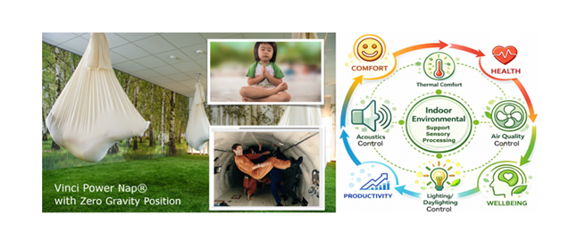
Picture 6. Vinci Power Nap® room with Zero Gravity Position and with indoor environmental elements influencing sensory processing and human well-being. Zero Gravity Position and its recreation on Earth in VPN. Picture of VPN green room at school: foto Pawel Pajot, source: own by author. Astronaut image used in illustration, source: (NASA Courses Neuro-Ophtalmology, Vestibular Physiology), child image: [781]. Illustration on right was generated using AI (ChatGPT 4.0.) and reviewed, corrected by the author.
Design frameworks such as Autism ASPECTSS™ [782] establish environmental guidelines to support sensory processing [778], while sensory research [9], [27], [665], [685], [686], [687], [695] underscores the impact of ambient stimuli like noise and others triggers on neurological function. These foundations suggest that targeted neuroarchitectural interventions, exemplified by the Vince Power Nap® pendulum’s harmonic with modulation of frequencies, could actively support autonomic regulation and sensory homeostasis in neurodiverse populations [688]. Furthermore, such
evidence-based design approaches highlight the need for interdisciplinary collaboration between architects, educators,
and healthcare professionals to optimize learning environments for children with diverse sensory needs.
10.5 The Need for RCT (Randomized Controlled Trial)
Randomized clinical trials are lacking, but preliminary data indicate promising directions in exploring mast cell overactivity, histamine metabolism, and anti-histamine diets in neuroimmunological, neuropsychiatric, and neurodevelopmental disorders including ASD, ADHD, PTSD, Alzheimer’s, and Parkinson’s, as well as the role of vitamin D, A, B6, B12, zinc, copper, selenium, H1/H2 blockers, DAO supplementation, and calming sensory environments in reducing excitotoxicity and excessive glutamatergic signaling [10], [12], [26]. Although there is a theoretical basis that increasing DAO activity may help alleviate ASD symptoms by reducing histamine, there is insufficient clinical evidence to confirm this. Additionally, the activity of the histamine-degrading enzyme diamine oxidase (DAO), necessary for adequate histamine breakdown, is significantly higher than the theoretical values provided in commercially available dietary supplements. Considering this, it is clear that improved supplements need to be developed to assist individuals with HIT, and thereby individuals with ASD and PTSD [455]. Further research is also needed to determine thresholds at which HVAC noise affects individuals with ASD and other sensory sensitivities. Studies should consider:
- The interaction between chronic low-level noise and neuroimmunological dysregulation.
- The cumulative impact of environmental factors, including noise, light, and temperature fluctuations [306], [625], [649], [650].
- Vinci Power Nap® helpful service to reduce stress, improve sleep and wellbeing for people wth ASD, ADHD, PTSD and their families [686].
- Interventions combining environmental modifications with dietary, pharmacological, and behavioral strategies. Since noise generated by HVAC systems is a modifiable environmental stressor that can significantly affect individuals with autism and other sensory sensitivities, incorporating building design, careful selection of HVAC systems, and individualized accommodations may improve quality of life and reduce behavioral and cognitive disturbances in these populations and how environmental stressors can amplifying histamine and glutamate loop (Picture 7).
Feedback loop: mast cells ↔ microglia ↔ neurons. In ASD, ADHD, and PTSD, a pathological loop develops: stress/sensory stimulus → mast cell activation → release of histamine and cytokines → microglial activation → neuroinflammation → lowered anxiety threshold → increased reactivity to stimuli → reactivation of mast cells → release of histamine and cytokines
This mechanism can explains sensory hypersensitivity, “fight or flight” responses without real danger, and difficulties with emotional regulation.
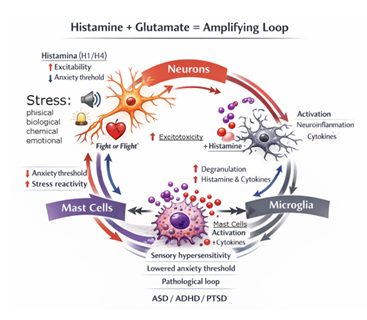
Picture 7: Showing that: Histamine + glutamate = amplifying loop: Mast cell activation: histamine (H1/H4) increases neuronal excitability, glutamate enhances microglial activation and mast cell degranulation, together they lower the anxiety threshold and increase stress reactivity. Illustration was generated using AI (ChatGPT 4.0.) and reviewed, corrected and completed by the author.
11. Discussion
This article does not mean that a cure for autism in humans has been found. Rather, it represents a step in research, pointing to potential regions that may be responsible for certain symptoms and suggesting mechanisms that could be targeted in future preventive and therapeutic interventions. Currently, there are no clinically effective therapies for ASD, ADHD, or PTSD. Nevertheless, positive pilot clinical studies using natural compounds with antioxidant and anti-inflammatory properties, which stabilize mast cells and thereby reduce histamine levels, offer hope [12], [26]. MCAS and HIT may contribute to symptoms frequently observed in ASD, ADHD, and PTSD [10]. Diagnosis is based on clinical symptom assessment, measurement of DAO enzyme levels, and observation of the patient’s response to a diet eliminating histamine-containing foods (or foods that trigger histamine release); implementing such a diet very often leads to rapid improvement in the patient’s condition [157]. Building on the author’s previous findings [10], [783], the present hypothesis expands this concept to a broader scale by proposing that chronic mast cell overactivation, together with impaired histamine metabolism, contributes to inflammatory processes, reduced GABA signaling, and excessive glutamatergic activity. These interconnected mechanisms are suggested to play a central role in the pathophysiology and symptom development of ASD, ADHD, and PTSD. This is often the result of complex interactions among neurotransmitters, genes, gut microbiome, immune system, and environmental factors. Preclinical data suggest that elements of interior architecture [666], exposure to stressors, Wi-Fi, ionizing radiation (X-rays, radiotherapy) (National Cancer Institute) [310], [643], as well as simultaneously consumed dietary components—for example, large amounts of ketchup (high histamine content), caffeine [90], casein, gluten, or wheat-containing products [784] —interact with these mechanisms, explaining the overlapping and amplifying nature of the symptoms [609]. Recognizing these relationships and eliminating them to prevent excessive activation of mast cells and microglia, as well as better understanding the mechanisms of HIT, may help limit neuroinflammatory processes [140] and open new therapeutic avenues [783]. Maintaining optimal levels of vitamin A (beta-carotene) and vitamin D (EDS CLINIC) [785] stabilizes mast cells, which, according to research, may also enhance antiviral immune responses, suppress retroviral infection [42], reduce histamine levels, and thus represent one of the key strategies in alleviating symptoms of MCAS, ASD, ADHD, and PTSD [284], [208], [209], [31].
Although ASD, PTSD, and ADHD differ in terms of developmental timing and clinical presentation, it looks that they share a common neuroimmunological pathway involving mast cell activation induced by chronic stress, neuroinflammation, dysregulation of histaminergic and glutamatergic signaling, and altered neuron–glia communication. Disorder-specific patterns reflect the timing and context of autonomic (immune–nervous) system programming: ASD has a prenatal origin (the system is “set” early), ADHD arises from a combination of developmental and environmental factors, and PTSD develops secondarily (the system is “reprogrammed” by trauma). A comprehensive review of ASD epidemiology in Arab countries by Newschaffer [786], categorized potential risk factors as: “1) maternal genetic predispositions, 2) environmental factors affecting the mother, 3) child genetic predispositions, and 4) environmental factors affecting the child, including absent or short breastfeeding, advanced maternal and paternal age, cesarean delivery, prenatal complications, vitamin D deficiency, exposure to pollutants, and heavy metals” [627]. According to [25] prenatal stress and inflammations [610] are associated with a higher risk of ASD in offspring [787], [610]. Mast cells in children with ASD have been observed to react disproportionately even to mild environmental triggers, where histamine can stimulate microglia, leading to dysfunction in neuronal connectivity and a low “fear threshold,” resulting in an exaggerated “fight-or-flight” response, which may be a key factor contributing to the pathogenesis of ASD and PTSD [788]. Future research on this hypothesis should focus on mast cell stabilization, DAO/HNMT genetics, histamine catabolism, dietary and environmental influences, and neuroimmunological interventions. Effective therapeutic strategies should consider individual differences in response to triggers, pharmacological, sensory, dietary, climatic, and environmental interventions in an integrated manner, forming a promising foundation for personalized therapies that provide relief to patients [789]. For example, clinical studies have shown that treatment with hydroxyzine (an H1 antihistamine) combined with a low-histamine diet led to significant overall improvement in individuals with intellectual disability [374], as well as treatment with H2 antihistamine could be a future therapeutic target of ADHD [790]. The simultaneous use of H1 and H2 receptor antagonists has shown positive effects not only in cardiac disease management [210], but also in the prevention and treatment of anaphylaxis [421], as well as in improving sleep architecture and behavior in children and adolescents with ASD [268], [270], [269], [258], and the antioxidant properties of H3 receptor antagonists have shown that these compounds may represent a promising therapeutic option in treating autism spectrum disorders (ASD) [258], [297]. These findings extend to other brain disorders in which microglia trigger inflammation, such as Alzheimer's disease [203], [209], schizophrenia [791], and BPSD [258], suggesting new strategies for treating neuroinflammation-related disorders [284]. Due to their ability to limit chemotaxis, H4 receptor antagonists are being studied in conditions such as atopic dermatitis, asthma, and chronic intestinal inflammation as potential anti-inflammatory and anti-allergic drugs [792], [793].
Considering histamine and the blood–brain barrier, drugs that suppress mast cell degranulation (e.g., luteolin [794], apigenin [795] and quercetin represent promising support in ASD therapy [796], [797]. Additionally, histamine receptor antagonists are presented as candidates for treating and preventing brain inflammation or edema [798], [214], as well as in improving post-stroke prognosis [101], [799], and in multiple sclerosis [137], which indicates that glial cells should be studied alongside mast cells as an interconnected network [138]. The importance of mast cells and the development of studies on their role in ASD is highlighted by [10] and can also be found in the later review article [30]. Variability in histamine metabolism among different individuals (e.g., levels of DAO, HNMT) depending on genetic, environmental, and dietary factors emphasizes the need for a personalized approach to the diagnosis and treatment of conditions that may be histamine-dependent, including symptoms of ASD, PTSD, ADHD, etc. [37]. Inflammatory states and immune dysregulation are key features characteristic of both neurodevelopmental and allergic disorders, suggesting overlapping pathophysiology. A 2025 study indicates that food, skin, and respiratory allergies are strongly associated with ADHD [800], [801]. Research from 2020 indicates that individuals with allergies have a 30–50% higher risk of ADHD and ASD [802]. Another study showed that children with ADHD exhibited rhinitis, eczema, significantly lower hemoglobin levels, higher IgE, and elevated eosinophil counts compared to controls, as well as microbiome dysbiosis and sleep disturbances [801]. A clinically important issue is the preparation of individuals with MCAS and HIT for surgical procedures, as invasive procedures and anesthesia are considered additional risk factors for anaphylaxis or adverse reactions “triggered by direct mast cell degranulation induced by mechanical irritation and tissue trauma, stress and pain, catheters, and anesthetic drugs during surgical procedures” [802]. Researchers propose the use of prophylactic antimediator therapy (PAT), including H1 and H2 histamine receptor antagonists [210], glucocorticoids, and benzodiazepines 1 hour prior to surgery, as this approach is not associated with adverse effects and reduces the likelihood of mast-cell mediator-dependent symptoms. Based on current evidence on the role of histamine in inflammatory, stress-related, and anaphylactoid reactions, it can be hypothesized that preoperative supplementation with diamine oxidase (DAO) and antihistamines may offer potential benefit in selected patients with HIT, MCAS [803], mastocytosis [804], or HIV infection [805], conditions that are associated with immune dysregulation and an increased prevalence of autoimmune comorbidities [806]. This proposed mechanism may involve a reduction in exogenous histamine load as well as a decrease in the overall histamine pool available to trigger systemic symptoms. However, this hypothesis remains speculative and requires confirmation through well-designed, controlled clinical trials and evidence-based clinical guidelines [774].
This review provides an understanding of these shared factors as potential mechanisms and their integration (interactions between mast cells, histamine, GABA/glutamate, and neuroinflammation) underlying the pathogenesis of ASD, ADHD [806], and PTSD [31], which may help explain comorbidities, overlapping symptoms, and suggest new therapies. The author emphasizes that this all suggests the existence of a shared etiology—in this case, the relationship between histamine levels and multiple symptoms—and calls for interdisciplinary research (including RCTs, genetics [811], environmental mast cell triggers [812], DAO, neonatal jaundice [813], D-dimers [814] and histamine levels) to test this hypothesis, taking into account individual variability. Conducting such studies will also allow for understanding the relationships between environmental elements and the human body, as well as applying safety engineering in the design of calming, human-centered spaces [807], particularly preventive spaces, with guidelines [782] for project elements—such as apartment layouts, selection of materials, colors, lighting, acoustics, shared spaces, and the outdoor environment [808] —which influence well-being and may reduce symptoms of ASD [809], ADHD, and PTSD, as exemplified by the pioneering neuroarchitecture system Vinci Power Nap® [665], [667], [668]. Recently, reports from the USA Department of Health and Human Services, working to find the cause of ASD, in September 2025 they announced indicated a correlation between prenatal acetaminophen use and later autism spectrum diagnoses in children (U.S. Department of Health and Human Services, 2025) [75]. However, the question arises whether the observed correlations are truly dependent on the medication itself, or rather on infections or inflammatory conditions during pregnancy (i.e., histamine release), the symptoms of which the mother attempted to alleviate with acetaminophen as an antipyretic and analgesic. The author’s previous discoveries and hypothesis [11], [13] were sent in emails dated 15.02.205 to the offices of Mr. Robert F. Kennedy Jr., Secretary General of the U.S. Department of Health, and emails dated 25.06.2025 with letter dated on 12 June 2025 to the offices of Mr. Robert F. Kennedy Jr. in MAHA and TeamKennedy, along with a link to the abstract of above hypothesis published on 19.05.2025 [10] to bring the attention of the Department while it was investigating the causes of autism spectrum disorder, with the intention of contributing to its ongoing efforts.
Picture 8: Letter to Mr. Robert F. Kennedy Jr., Secretary General of the U.S. Department of Health
12. Conclusion
The findings suggest the existence of a pathophysiological continuity between earlier exposures or biological events and later clinical manifestations. The collected data indicate that ASD and related neuropsychiatric conditions, such as ADHD and PTSD, result from a complex interaction between the nervous system, the immune system, and the environment. Analysis of the available data and literature suggests that disorders associated with chronic mast cell hyperactivation—and consequently with histamine metabolism, the related dysregulation of the glutamate–GABA balance, and chronic neuroinflammation—may play a key role in the pathophysiology of ASD, ADHD, PTSD, as well as other neuroimmunological disorders in which this pathological loop develops. Accordingly, stabilization of mast cell activation, reduction of histamine levels, and modulation of neuroinflammatory pathways represent promising preventive and therapeutic strategies that may support cognitive functions, emotional regulation, and improve the overall quality of life of individuals affected by neurodegenerative disorders and gastrointestinal symptoms. In individuals with ASD, impairments in the enzymatic degradation of histamine (DAO, HNMT) are frequently observed, which may lead to excessive histamine accumulation in the body and secondary mast cell activation. A low-histamine diet, supplementation with the DAO enzyme, vitamins D3, A, B6, B9, and B12, as well as minerals such as zinc, selenium, and copper, acemannan, together with appropriate sensory interventions, show potential in supporting the management of symptoms related to neuronal hyper-excitability, heightened sensitivity, and maladaptive stress responses.
Within a personalized medicine framework that accounts for individual genetic variability in histamine metabolism pathways, selected natural bioactive compounds—such as quercetin, apigenin, and luteolin—black seed oil, as well as certain antihistamine medications (H1, H2, and H3 receptor antagonists), may exert mast cell–stabilizing effects, limit neuroinflammatory processes, and support blood–brain barrier integrity, potentially providing therapeutic benefits for selected patients with ASD, ADHD, and PTSD; however, this requires further validation in controlled studies. The environment also plays a significant role in modulating the aforementioned processes. Adverse environmental factors, stress, pollution, intrusive technologies and sensory overstimulation may exert epigenetic effects on mast cell hyperreactivity, thereby increasing the risk of ASD development (Marzi et l. 2025). Biological, physical, and chemical factors—such as molds, fungi, bacteria; toxins, poisons, and medications; cold and noise (including low-frequency noise emitted by air-conditioning systems), vibrations, mechanical pressure (e.g., elastic bands in clothing), air pollution, heavy metals, microplastics, ambient temperature, excessive light exposure, and electromagnetic radiation—may exacerbate sensory stress (similarly to prenatal stress, emotional stress, and trauma) and induce IgE-independent mast cell degranulation. This can leads to increased levels of histamine and pro-inflammatory cytokines, and consequently to infection-like symptoms, neonatal jaundice, blood–brain barrier permeability, microglial activation, elevated glutamate levels, neuroinflammation, lowering of the anxiety threshold, heightened stimulus reactivity, and further mast cell activation.
In the context of sensory hypersensitivity, “fight-or-flight” responses in the absence of real threat, as well as sleep disturbances and difficulties with emotional regulation, suggest that appropriately designed, supportive surroundings characterized by low noise levels, comfortable temperatures, access to green spaces, biophilic design features, and a strong sense of safety may promote recovery and well-being. Such environments can facilitate neuroregenerative processes, enhance emotional regulation, and provide restorative benefits for both affected individuals and their families. Although ASD, ADHD, and PTSD differ in their timing of onset and clinical presentation, at the cellular level they activate similar neuroimmunological pathways associated with chronic stress, neuroinflammation, and excessive neuronal excitability. Prolonged activation of mast cells and histamine release may influence inflammatory processes that could potentially shorten lifespan, suggesting a link between mast cell regulation and longevity. Regulation of mast cells and histamine may represent a key mechanism affecting aging processes and lifespan. Despite the growing number of descriptive studies and case reports, randomized interdisciplinary clinical trials (RCTs) are needed to confirm the proposed hypothesis and the effectiveness of the suggested interventions. Understanding the converging mechanisms of mast cell activation, histamine signaling, neurotransmitter imbalance (GABA/glutamate), and neuroinflammation will enable more accurate assessment of overlapping symptoms and the identification of novel therapeutic targets. Future research should focus on mast cell activity, genetic determinants of DAO/HNMT enzymes, the impact of the sensory environment, and integrated neuroimmunological therapy models encompassing pharmacological, dietary, behavioral, and architectural components, in order to evaluate their effects on the prevention or progression of many diseases.
A holistic approach integrating knowledge from cellular biology, neurochemistry, neuroarchitecture, and safety engineering opens new perspectives for the personalized medicine of the future—aimed not only at symptom reduction but also at improving the quality of life of individuals with ASD, PTSD, ADHD, MCAS, HIT, and other disorders, including Alzheimer’s and Parkinson’s diseases. Increasing evidence suggests that, in many cases, the underlying basis of symptoms may be neurochemical rather than exclusively psychological. This work constitutes a biological bridge between immunology, neurobiology, neuroarchitecture, developmental psychiatry, sleep, longevity and personalized medicine.
Acknowledgements
- Special thanks are extended to the Special Education and Care Center in Piła, Poland (Specjalny Ośrodek Szkolno-Wychowawczy im. Marii Grzegorzewskiej w Pile), including its Directors and therapists, especially Karolina Kosterka-Dorsz, for their support and active participation in the pilot project. Their openness and collaboration have been invaluable in bridging theory with practice. Informed consent was obtained from all subjects involved in the study.
Conflict of interest statement
Author is also the creator and researcher of Vinci Power Nap® neuroarchitecture pioneering system.
GenAI Disclosure Statement
ChatGPT 4.0 has strictly been used for grammar review, writing clarity in some sections of this paper and help with infographic. No part of the substantive content, arguments, or conclusions were generated by AI, they are solely the work of the author. Chosen illustrations was generated using AI ChatGPT 4.0., reviewed, corrected and completed by the author.
List of Abbreviations
AA – Arachidonic Acid
ACLF – Acute-on-Chronic Liver Failure
ACLD – Advanced Chronic Liver Disease
ADHD – Attention-Deficit/Hyperactivity Disorder
ADDM – Autism and Developmental Disabilities Monitoring
ALDH7A1 – Aldehyde Dehydrogenase 7 Family Member A1
AMPA – α-amino-3-hydroxy-5-methyl-4-isoxazolepropionic acid receptor (ionotropic glutamate receptor)
ASD – Autism Spectrum Disorder
BBB – Blood–Brain Barrier
BCM7 – Peptide beta-casomorphin-7
BCSFB – Blood–Cerebrospinal Fluid Barrier
BLB – Blood–Labyrinth Barrier
BNB – Blood–Nerve Barrier
CDC – Centers for Disease Control and Prevention
CGRP – Calcitonin Gene-Related Peptide
CLP – Cecal Ligation and Puncture
COMT – Catechol-O-Methyltransferase
COPD – Chronic Obstructive Pulmonary Disease
CRF – Corticotropin-Releasing Factor
CRH/CRF – Corticotropin-Releasing Hormone / Factor
CRP – C-Reactive Protein
CNS – Central Nervous System
DAO – Diamine Oxidase
DHA – Docosahexaenoic Acid
DIC – Disseminated Intravascular Coagulation
DNMT – DNA Methyltransferases
ECL – Enterochromaffin-like Cells
EBV – Epstein-Barr Virus
ESR – Erythrocyte Sedimentation Rate
GABA – Gamma-Aminobutyric Acid
GERD – Gastroesophageal Reflux Disease
GGT – Gamma-Glutamyl Transferase
HNMT – Histamine N-Methyltransferase
HDC – Enzyme Histidine Decarboxylase
HCl – Hydrochloric Acid
HPA axis – Hypothalamic-Pituitary-Adrenal Axis
HIT – Histamine Intolerance
IL-1β – Interleukin-1 Beta (silnie prozapalna cytokina)
IL-6 – Interleukin-6 (cytokina o działaniu dwoistym: ostra faza vs przewlekłe zapalenie)
LTP – Long-Term Potentiation
MAOB – Monoamine Oxidase B
MCs – Mast Cells
MCAS – Mast Cell Activation Syndrome / Mastocytosis
MSG – Monosodium Glutamate
NMDA – N-Methyl-D-Aspartate Receptor (jonotropowy receptor glutaminianu)
NT – Neurotensin
NSAIDs – Nonsteroidal Anti-Inflammatory Drugs
OXTR – Oxytocin Receptor
POTS – Postural Orthostatic Tachycardia Syndrome
PTH – Parathyroid Hormone
PMDD – Premenstrual Dysphoric Disorder
PMS – Premenstrual Syndrome
RCT – Randomized Clinical Trials
SAH – S-adenosylhomocysteine
SAM – S-adenosylmethionine
SCF – Stem Cell Factor
SCH – Schizophrenia
SIBO – Small Intestinal Bacterial Overgrowth
SM – Systemic Mastocytosis
SIDS – Sudden Infant Death Syndrome
SIRS – Systemic Inflammatory Response Syndrome
TGF-β – Transforming Growth Factor Beta (cytokina regulacyjna / immunosupresyjna)
TNF-α – Tumor Necrosis Factor Alpha
VPN - Vinci Power Nap®
WHO – World Health Organization
5-HT – Serotonin
References
- CDC (2025). https://www.cdc.gov/autism/data-research/?utm_source=chatgpt.com
- Shaw K A, Williams S, Patrick M E, et al. Prevalence and early identification of autism spectrum disorder among children aged 4 and 8 years—Autism and Developmental Disabilities Monitoring Network, 16 sites, United States, 2022. MMWR Surveillance Summaries 74 (2025): 1-22.
- CDC. Prevalence of autism spectrum disorder. Morbidity and Mortality Weekly Report 69 (2020): 1-12.
- Ayano G, Demelash S, et al. The global prevalence of attention deficit hyperactivity disorder in children and adolescents: An umbrella review of meta-analyses. Journal of Affective Disorders (2023).
- Zablotsky B, Black LI, Maenner MJ, et al. Prevalence and Trends of Developmental Disabilities among Children in the United States: 2009–2017. Pediatrics 144 (2019): e20190811.
- Evans G W. The environment of childhood poverty. The American psychologist 59 (2004): 77-92.
- Kessler R C, Berglund P, et al. Lifetime prevalence and age-of-onset distributions of DSM-IV disorders in the National Comorbidity Survey replication. Archives of General Psychiatry 62 (2005): 593-602.
- Mental Health America. What are the symptoms of PTSD? https://screening-mhanational-org.translate.goog/content/what-are-symptoms-ptsd/?_x_tr_sl=en&_x_tr_tl=pl&_x_tr_hl=pl&_x_tr_pto=sge#:~:text=You%20may%20be%20easily%20startled,Avoiding%20reminders%20of%20the%20trauma
- Filcek M, Vats M. Reduction of Stress and Jet Lag, Improvement of Well-Being, Sleep Quality and Body-Mind Regeneration by Vinci Power Nap® Neuroarchitecture System: Innovative Solution for Leaders, Employees, Travelers. IntechOpen. Book: Sleep or Awake? (2023).
- Filcek M. Discovery of Pathophysiological Mechanism Underlying Symptoms of ASD and PTSD: The Role of Mast Cell Hyperactivation and Histamine Metabolic Imbalance. Peeref publication (2025).
- Filcek M, Vats M, Skrzyniarz-Plutecka A. Discovery of the Mechanism of COVID-19, SIRS and Sepsis: Mast Cells and Histamine Storm as Overlooked Aspects in COVID-19 and in Ventilated Patients—Potential Role of Antihistamines MAR Pulmonology 3 (2021): 5. https://drive.google.com/file/d/16w5eWwKK4ppPgGX-zXXVEH4Hhh_O3G3Y/view ; https://www.medicalandresearch.com/uploads/current_issues/1696542294_1783152566.pdf
- Theoharides T C, et al. Mast cells, brain inflammation and autism. European Journal of Pharmacology 778 (2016): 96-102
- Filcek M, Vats M, Skrzyniarz-Plutecka A. “COVID-19 and Antihistaminic Medicines - Improving the Health of Millions by Strategically Disseminating a "Simple but Potential Solution"” MAR Pulmonology 3 (2021): 1. https://drive.google.com/file/d/1o-BCL7UJ0wwLSZ6BDQnOIi8KkHrtpmn2/view ; https://www.medicalandresearch.com/uploads/current_issues/451827006_1782991949.pdf
- Albrecht M, Dittrich A M. Expression and function of histamine and its receptors in atopic dermatitis. Molecular and Cellular Pediatrics 2 (2015): 16.
- Thangam E B, Jemima E A, et al. The role of histamine and histamine receptors in mast cell-mediated allergy and inflammation: The hunt for new therapeutic targets. Frontiers in Immunology 9 (2018).
- Panula P, Chazot P L, et al. International Union of Basic and Clinical Pharmacology. XCVIII. Histamine receptors. Pharmacological Reviews 67 (2015): 601-655.
- Borecki M, Jałocha K, et al. Histamine Intolerance on Diagnosis and Treatment. Medical Science 29 (2025): e85ms3580.
- El-Ansary A. GABA and Glutamate Imbalance in Autism and Their Reversal as Novel Hypothesis for Effective Treatment Strategy (2020).
- Hamed N O, Al-Ayadhi L, et al. Understanding the roles of glutamine synthetase, glutaminase, and glutamate decarboxylase autoantibodies in imbalanced excitatory/inhibitory neurotransmission as etiological mechanisms of autism. Psychiatry and clinical neurosciences 72 (2018): 362-373.
- Arrang J M, Garbarg M, et al. Highly potent and selective ligands for histamine H3-receptors. Nature 327 (1987): 117-123.
- Arrang J M, Garbarg M, Schwartz J C. Auto-inhibition of brain histamine release mediated by a novel class (H3) of histamine receptor. Nature 327 (1987): 117-123.
- El-Ansary A, Al-Ayadhi L. GABAergic/glutamatergic imbalance relative to excessive neuroinflammation in autism spectrum disorders. Journal of neuroinflammation, 11 (2014): 189.
- Theoharides T C, Alysandratos K D, et al. Mast cells and inflammation. Biochimica et biophysica acta 1822 (2012): 21-33.
- Theoharides T C, Angelidou A, et al. Mast cell activation and autism. Biochimica et biophysica acta 1822 (2012).
- Theoharides T C, Kavalioti M, Tsilioni I. Mast Cells, Stress, Fear and Autism Spectrum Disorder. International Journal of Molecular Sciences 20 (2019): 3611.
- Theoharides T C. Danger Signals and Inflammation. Clinical Therapeutics 38 (2016): 996-999.
- Filcek M. Oddech Breathe with Vinci Power Nap - harmonize your body and mind in the Sleep Cafe. A neuroarchitectural system for synchronizing sleep, breathing, heartbeat, and brainwaves that reduces stress, mast cell activation stimuli, and histamine. Chapter: 22 in book: Oddech, Edition: Mira Rządzka, Publisher: Impuls (2025).
- Pytka M, Dalmata W, Należna P. The neuroinflammation origin of autism spectrum disorder (ASD) – a literature review. Journal of Education, Health and Sport 84 (2025): 39926.
- Theoharides T C. Ways to Address Perinatal Mast Cell Activation and Focal Brain Inflammation, including Response to SARS-CoV-2, in Autism Spectrum Disorder. Journal of Personalized Medicine 11 (2021): 860.
- Priego-González L, Pardo P P, Redegeld F. The role of mast cells in autism spectrum disorder. Neuroscience and Biobehavioral Reviews 176 (2025): 106263.
- Kempuraj D, Selvakumar G P, et al. Mast Cell Activation in Brain Injury, Stress, and Post-traumatic Stress Disorder and Alzheimer's Disease Pathogenesis. Frontiers in neuroscience 11 (2017): 703.
- Tamasi J, Kalabay L. Spectrum, time course, stages, and a proposal for the diagnosis of histamine intolerance in general practice: A nonrandomized, quasi-experimental study. Journal of Clinical Medicine 14 (2025): 311.
- Renkielska D. Komórki tuczne w przewlekłych zmianach okołowierzchołkowych szczęk. Gdański Uniwersytet Medyczny (2014).
- Sagües-Sesé E, García-Casares N, Álvarez-Twose I. Cognitive, neuropsychiatric and neurological alterations in mastocytosis: A systematic review. Clinical and Translational Allergy 13 (2023): e12319.
- Zasowska-Nowak A, Ciałkowska-Rysz A. Systemic mastocytosis – a complex problem of symptomatic treatment. Medycyna Paliatywna/Palliative Medicine 12 (2020): 47-55.
- Persico A M, Bourgeron T. Searching for ways out of the autism maze: genetic, epigenetic and environmental clues. Trends in neurosciences 29 (2006): 349-358.
- Blasco-Fontecilla H, Bella-Fernández M, Wang P, et al. Prevalence and Clinical Picture of Diamine Oxidase Gene Variants in Children and Adolescents with Attention Deficit Hyperactivity Disorder: A Pilot Study. Journal of clinical medicine 13 (2024): 1659.
- Chung S, Hong J P, Yoo H K. Association of the DAO and DAOA gene polymorphisms with autism spectrum disorders in boys in Korea: a preliminary study. Psychiatry research 153 (2007): 179-182.
- Kadiyska T, Tourtourikov I, et al. Impact of AOC1 and HNMT variants on the therapeutic outcomes of a histamine reducing diet in autism spectrum disorder. Journal of Molecular Neuroscience 75 (2025): 105.
- Theoharides - video: Role of Mast Cells in Inflammation of the Brain and Autism Spectrum Disorder Professor Theoharides. [www.youtube.com/watch?v=rW2K_WPUQTA]
- Theoharides T C, Tsilioni I, Ren H. Recent advances in our understanding of mast cell activation—or should it be mast cell mediator disorders? Expert Review of Clinical Immunology 15 (2019).
- Song S T, Wu M L, Zhang H J, Su X, Wang J H. Mast cell activation triggered by retrovirus promotes acute viral infection. Frontiers in Microbiology 13 (2022): 798660.
- Stone K D, Prussin C, Metcalfe D D. IgE, mast cells, basophils, and eosinophils. Journal of Allergy and Clinical Immunology 125 (2010): 73-80.
- Dong H, Zhang X, Qian Y. Mast cells and neuroinflammation. Medical science monitor basic research 20 (2014): 200-206.
- Silver R, Silverman A J. Mast cells in the brain: Evidence and functional significance. Trends in Neurosciences 31 (2008): 358-365.
- Pang X, et al. Mast cell degranulation and release of histamine in the rat brain induced by compound 48/80. Journal of Neurochemistry 67 (1996):1098-1105.
- Conti P, Kempuraj D. Important role of mast cells in multiple sclerosis. Mult Scler Relat Disord 5 (2016): 77-80.
- Theoharides T C, Tsilioni I, Ren H. Recent advances in our understanding of mast cell activation—or should it be mast cell mediator disorders? Expert Review of Clinical Immunology, 15 (2019).
- Galli S J, Gaudenzio N, Tsai M. Mast Cells in Inflammation and Disease: Recent Progress and Ongoing Concerns. Annual review of immunology 38 (2020): 49-77.
- Sumantri S, Rengganis I. Immunological dysfunction and mast cell activation syndrome in long COVID. Asia Pacific Allergy 13 (2023): 50-53.
- Yu M, Song X T, et al. The Emerging Role of Mast Cells in Response to Fungal Infection. Frontiers in Immunology 12 (2021): 688659.
- Özdemir Ö, Kasımoğlu G, Bak A, et al. Mast cell activation syndrome: An up-to-date review of literature. World Journal of Clinical Pediatrics 13 (2024): 92813.
- Sánchez-Borges M, Caballero-Fonseca F, Capriles-Hulett A, et al. Hypersensitivity Reactions to Nonsteroidal Anti-Inflammatory Drugs: An Update. Pharmaceuticals 3 (2010): 10-18.
- Schmidt-Rondon E, Wang Z, Malkmus SA, et al. Effects of opioid and nonopioid analgesics on canine wheal formation and cultured human mast cell degranulation. Toxicology and Applied Pharmacology 338 (2018): 54-64.
- Fukuoka Y, Schwartz DL, Ward BR. Activation of human skin mast cells by vancomycin via MrgX2: Comparison to the effects of brimonidine. Journal of Immunology 206 (2021): 23.10.
- Lide B, Mcguire S, Liu H, et al. Mast cell activation syndrome-anesthetic challenges in two different clinical scenarios. Journal of Biomedical Research 36 (2022): 435-439.
- Mirone C, Preziosi D, Mascheri A, et al. Identification of risk factors of severe hypersensitivity reactions in general anaesthesia. Clinical and Molecular Allergy 13 (2015): 11.
- Mertes PM, Malinovsky JM, Jouffroy L, et al. Reducing the risk of anaphylaxis during anesthesia: 2011 updated guidelines for clinical practice. Journal of Investigational Allergology and Clinical Immunology 21 (2011): 442-453.
- Jiang W, Hu S, Che D, et al. A mast-cell-specific receptor mediates Iopamidol induced immediate IgE-independent anaphylactoid reactions. International Immunopharmacology 75 (2019): 105800.
- Baldo BA, Pham NH. Opioid toxicity: histamine, hypersensitivity, and MRGPRX2. Archives of Toxicology 97 (2023): 359-375.
- Rama TA, Castells M. Triggers of Anaphylaxis in Mastocytosis Patients: Evidence of the Current Drug-Avoidance Recommendation. Current Treatment Options in Allergy 10 (2023): 442-457.
- Gargano D, Appanna R, Santonicola A, et al. Food Allergy and Intolerance: A Narrative Review on Nutritional Concerns. Nutrients 13 (2021): 1638.
- Schedle A, Samorapoompichit P, Füreder W, et al. Metal ion-induced toxic histamine release from human basophils and mast cells. Journal of Biomedical Materials Research 39 (1998): 560-567.
- Walczak-Drzewiecka A, Wyczólkowska J, Dastych J. Environmentally relevant metal and transition metal ions enhance Fc epsilon RI-mediated mast cell activation. Environmental Health Perspectives 111 (2003): 708-713.
- Visalli G, Facciolà A, Pruiti Ciarello M, et al. Acute and Sub-Chronic Effects of Microplastics (3 and 10 µm) on the Human Intestinal Cells HT-29. International Journal of Environmental Research and Public Health 18 (2021): 5833.
- Khan A, Jia Z. Recent insights into uptake, toxicity, and molecular targets of microplastics and nanoplastics relevant to human health impacts. iScience 26 (2023).
- Yamanaka-Takaichi M, et al. Stress and Nasal Allergy: Corticotropin-Releasing Hormone Stimulates Mast Cell Degranulation and Proliferation in Human Nasal Mucosa. International Journal of Molecular Sciences 22 (2021): 2773.
- El-Ansary A, Al-Dbass A, Qasem H. Neurotoxins and Autism (2018).
- Geier DA, Geier MR. A case-control study of serious autoimmune adverse events following hepatitis B immunization. Autoimmunity 38 (2005): 295-301.
- Geier MR, Geier DA. A case-series of adverse events, positive re-challenge of symptoms, and events in identical twins following hepatitis B vaccination: analysis of the Vaccine Adverse Event Reporting System (VAERS) database and literature review. Clinical and Experimental Rheumatology 22 (2004): 749-755.
- Heavy metals - video. https://www.instagram.com/reel/DI4shgZy8W_/?igsh=YTNlZ3ZybngwOHBu
- RFKennedy - videos. (https://www.instagram.com/reel/DG78kDhNhJk/?igsh=MW5ubmV0aWExdjF5eQ==); (https://www.instagram.com/reel/DJWcA4Oi5ui/?igsh=ajQ0ZGszbjRleWR3)
- Thimerosal-containing vaccines and neurodevelopmental outcomes. Review of the evidence. https://cdn.factcheck.org/UploadedFiles/Thimerosal-Containing-Vaccines-Summary-of-Evidence-508.pdf.
- Thimerosal and Vaccines. https://www.fda.gov/vaccines-blood-biologics/safety-availability-biologics/thimerosal-and-vaccines
- S. Department of Health and Human Services. President Trump, Secretary Kennedy announce bold actions to tackle autism epidemic (2025). https://www.hhs.gov/press-room/hhs-trump-kennedy-autism-initiatives-leucovorin-tylenol-research-2025.html
- McSweeney SM, Christou EAA, et al. Physical urticaria: Clinical features, pathogenesis, diagnostic work-up, and management. Journal of the American Academy of Dermatology 89 (2023): 324-337.
- Müller K, Meineke V. Radiation-induced mast cell mediators differentially modulate chemokine release from dermal fibroblasts. Journal of Dermatological Science 61 (2011): 199-205.
- Kaplan AP, Beaven MA. In vivo studies of the pathogenesis of cold urticaria, cholinergic urticaria, and vibration-induced swelling. Journal of Investigative Dermatology 67 (1976): 327-332.
- Gausche-Hill M, Fuchs S, Yamamoto L. APLS: The Pediatric Emergency Medicine Resource. Jones & Bartlett Learning (2004): 69.
- Khan BQ, Kemp SF. Pathophysiology of anaphylaxis. Current Opinion in Allergy and Clinical Immunology 11 (2011): 319-325.
- Theoharides TC. The impact of psychological stress on mast cells. Annals of Allergy, Asthma & Immunology 125 (2020): 388-392.
- Santos J, Yang PC, Söderholm JD, et al. Role of mast cells in chronic stress-induced colonic epithelial barrier dysfunction in the rat. Gut 48 (2001): 630-636.
- Eutamene H, Theodorou V, Fioramonti J, et al. Acute stress modulates the histamine content of mast cells in the gastrointestinal tract through interleukin-1 and corticotropin-releasing factor release in rats. Journal of Physiology 553 (2003): 959-966.
- Kilinc E, Dagistan Y, Tore F. Mast Cell Degranulation Mediates Compound 48/80-Induced Meningeal Vasodilatation Underlying Migraine Pain. Clinical and Experimental Health Sciences 8 (2018): 196+. https://link.gale.com/apps/doc/A569683658/AONE?u=anon~3f95d6ae&sid=googleScholar&xid=0c0d5967
- Bódis J, Tinneberg HR, Schwarz H, et al. The effect of histamine on progesterone and estradiol secretion of human granulosa cells in serum-free culture. Gynecological Endocrinology 7 (1993): 235-239.
- Schirmer B, Neumann D. The Function of the Histamine H4 Receptor in Inflammatory and Inflammation-Associated Diseases of the Gut. International Journal of Molecular Sciences 22 (2021): 6116.
- Smolinska S, Winiarska E, Globinska A, et al. Histamine: A Mediator of Intestinal Disorders—A Review. Metabolites 12 (2022): 895.
- Zhao Y, et al. Histamine Intolerance—A Kind of Pseudoallergic Reaction. Biomolecules 12 (2022): 454.
- Hrubisko M, Danis R, Huorka M, et al. Histamine Intolerance-The More We Know the Less We Know. A Review. Nutrients 13 (2021): 2228.
- John J, Kodama T, Siegel JM. Caffeine promotes glutamate and histamine release in the posterior hypothalamus. American Journal of Physiology: Regulatory, Integrative and Comparative Physiology 307 (2014): R704-R710.
- Moon TC, Befus AD, Kulka M. Mast cell mediators: their differential release and the secretory pathways involved. Frontiers in Immunology 5 (2014): 569.
- Schäfer T, Starkl P, Allard C, et al. A granular variant of CD63 is a regulator of repeated human mast cell degranulation. Allergy 65 (2010): 1242-1255.
- Provensi G, Costa A, et al. Brain histamine modulates recognition memory: Possible implications in major cognitive disorders. British Journal of Pharmacology 177 (2020): 539-556.
- Reber LL, Hernandez JD, Galli SJ. The pathophysiology of anaphylaxis. Journal of Allergy and Clinical Immunology 140 (2017): 335-348.
- Eklund KK, Humphries DE, Xia Z, et al. Glucocorticoids inhibit the cytokine-induced proliferation of mast cells, the high affinity IgE receptor-mediated expression of TNF-alpha, and the IL-10-induced expression of chymases. Journal of Immunology 158 (1997): 4373-4380.
- Alotiby A. Immunology of Stress: A Review Article. Journal of Clinical Medicine 13 (2024): 6394.
- Tsai M, Valent P, Galli SJ. KIT as a master regulator of the mast cell lineage. Journal of Allergy and Clinical Immunology 149 (2022): 1845-1854.
- Metcalfe DD. Mast cells and mastocytosis. Blood 112 (2008): 946-956.
- Seeley EJ, Sutherland RE, Kim SS, et al. Systemic mast cell degranulation increases mortality during polymicrobial septic peritonitis in mice. Journal of Leukocyte Biology 90 (2011): 591-597.
- Ramos L, Pena G, Cai B, et al. Mast cell stabilization improves survival by preventing apoptosis in sepsis. Journal of Immunology 185 (2010): 709-716.
- Blasco MP, Chauhan A, Honarpisheh P, et al. Age-dependent involvement of gut mast cells and histamine in post-stroke inflammation. Journal of Neuroinflammation 17 (2020): 160.
- Żelechowska P, Agier J, Kozłowska E, et al. Mast cells participate in chronic low-grade inflammation within adipose tissue. Obesity Reviews 1 (2018): 686-697.
- Zhang Z, Kurashima Y. Two Sides of the Coin: Mast Cells as a Key Regulator of Allergy and Acute/Chronic Inflammation. Cells 10 (2021): 1615.
- Mazurek T. Proinflammatory capacity of adipose tissue – new insights in the pathophysiology of atherosclerosis. Kardiologia Polska 67 (2009): 1119-1124.
- Wensveen FM, Valentić S, et al. The "Big Bang" in obese fat: Events initiating obesity-induced adipose tissue inflammation. European Journal of Immunology 45 (2015): 2446-2456.
- Hotamisligil GS. Inflammation and metabolic disorders. Nature 444 (2006).
- Żelechowska P, Agier J, et al. Leptin stimulates tissue rat mast cell pro-inflammatory activity and migratory response. Inflammation Research 67 (2018): 789-799.
- Parrella E, Porrini V, Benarese M, et al. The Role of Mast Cells in Stroke. Cells 8 (2019): 437.
- Lumeng CN, Saltiel AR. Inflammatory links between obesity and metabolic disease. Journal of Clinical Investigation 121 (2011): 2111-2117.
- Khouma A, Moeini MM, Plamondon J, et al. Histaminergic regulation of food intake. Frontiers in Endocrinology 14 (2023): 1202089.
- Grund VR, Goldberg ND, Hunninghake DB. Histamine receptors in adipose tissue: involvement of cyclic adenosine monophosphate and the H2-receptor in the lipolytic response to histamine in isolated canine fat cells. Journal of Pharmacology and Experimental Therapeutics 195 (1975): 176-184.
- Divoux A, Moutel S, et al. Mast cells in human adipose tissue: link with morbid obesity, inflammatory status, and diabetes. Journal of Clinical Endocrinology and Metabolism 97 (2012): E1677-E1685.
- Geller V, Dominiak M. Systemic immune-inflammatory and response indices as quantitative biomarkers for obesity. Obesity Facts 18 (2025): 572-573.
- Shi MA, Shi GP. Different roles of mast cells in obesity and diabetes: lessons from experimental animals and humans. Frontiers in Immunology 3 (2012): 7.
- Sismanopoulos N, Delivanis DA, Mavrommati D, et al. Do mast cells link obesity and asthma? Allergy 68 (2013): 8-15.
- Sammels O, Karjalainen L, Dahlgren J, et al. Autism Spectrum Disorder and Obesity in Children: A Systematic Review and Meta-Analysis. Obesity Facts 15 (2022): 305-320.
- Curtin C, Anderson SE, Must A, et al. The prevalence of obesity in children with autism: a secondary data analysis using nationally representative data from the National Survey of Children's Health. BMC Pediatrics 10 (2010): 11.
- Curtin C, Jojic M, Bandini LG. Obesity in children with autism spectrum disorder. Harvard Review of Psychiatry 22 (2014): 93-103.
- Tye C, Runicles AK, Whitehouse AJO, et al. Characterizing the interplay between autism spectrum disorder and comorbid medical conditions: An integrative review. Frontiers in Psychiatry 9 (2019): 751.
- Plum T, Feyerabend TB, Rodewald HR. Beyond classical immunity: Mast cells as signal converters between tissues and neurons. Immunity 57 (2024): 2723-2736.
- Alizadeh A, Dyck SM, Karimi-Abdolrezaee S. Myelin damage and repair in pathologic CNS: Challenges and prospects. Frontiers in Molecular Neuroscience 8 (2015): 35.
- Yang J, et al. Laparotomy-induced peripheral inflammation activates NR2B receptors on brain mast cells and results in neuroinflammation in a vagus nerve-dependent manner. Frontiers in Cellular Neuroscience 16 (2022): 771156.
- Grootens J, et al. Deciphering the differentiation trajectory from hematopoietic stem cells to mast cells. Blood Advances 2 (2018): 2273-2281.
- Ribatti D, d'Amati A. Hematopoiesis and mast cell development. International Journal of Molecular Sciences 24 (2023).
- Kim DK, et al. Mastocytosis-derived extracellular vesicles deliver miR-23a and miR-30a into pre-osteoblasts and prevent osteoblastogenesis and bone formation. Nature Communications 12 (2021): 2527.
- Sacks D, Baxter B, et al. Multisociety Consensus Quality Improvement Revised Consensus Statement for Endovascular Therapy of Acute Ischemic Stroke. International Journal of Stroke 13 (2018): 612-632.
- Johnson D, Krenger W. Interactions of mast cells with the nervous system--recent advances. Neurochemical Research 17 (1992): 939-951.
- Kempuraj D, Mentor S, et al. Mast Cells in Stress, Pain, Blood-Brain Barrier, Neuroinflammation and Alzheimer’s Disease. Frontiers in Cellular Neuroscience 13 (2019): 54.
- Rocha SM, Saraiva T, et al. Histamine induces microglia activation and dopaminergic neuronal toxicity via H1 receptor activation. Journal of Neuroinflammation 13 (2016): 137.
- Strider JW, Masterson CG, Durham PL. Treatment of mast cells with carbon dioxide suppresses degranulation via a novel mechanism involving repression of increased intracellular calcium levels. Allergy 66 (2011): 341-350.
- Liu ZQ, Li XX, et al. Vitamin D contributes to mast cell stabilization. Allergy 72 (2017): 1184-1192.
- Mehrani Y, Morovati S, et al. Vitamin D Influences the Activity of Mast Cells in Allergic Manifestations and Potentiates Their Effector Functions against Pathogens. Cells 12 (2023): 2271.
- El-Ansary A, Qasem H. Vitamin D as nutritional strategy to treat autism. Nutrition & Food Technology: Open Access 1 (2018).
- Jin Y, Silverman AJ, Vannucci SJ. Mast cells are early responders after hypoxia–ischemia in immature rat brain. Stroke 40 (2009): 3107-3112.
- Rönnbäck C, Hansson E. The Importance and Control of Low-Grade Inflammation Due to Damage of Cellular Barrier Systems That May Lead to Systemic Inflammation. Frontiers in Neurology 10 (2019): 533.
- Vakilzadeh G, Martinez-Cerdeño V. Pathology and Astrocytes in Autism. Neuropsychiatric Disease and Treatment 19 (2023): 841-850.
- Kim DY, Hong GU, Ro JY. Signal pathways in astrocytes activated by cross-talk between astrocytes and mast cells through CD40-CD40L. Journal of Neuroinflammation 8 (2011): 25.
- Skaper SD, Facci L, Giusti P. Mast cells, glia and neuroinflammation: partners in crime? Immunology 141 (2013): 314-327.
- Karolinska Institute. Karolinska Institute (2013). ki.se
- Hendriksen E, van Bergeijk D, Oosting RS, et al. Mast cells in neuroinflammation and brain disorders. Neuroscience and Biobehavioral Reviews 79 (2017): 119-133.
- Barger G, Dale HH. 4-β-aminoethylglyoxaline (β-iminazolylethylamine) and the other active principles of ergot. Journal of the Chemical Society, Transactions 97 (1910): 2592-2595.
- Dale HH. The physiological action of histamine. Journal of Physiology 51 (1920): 1-18.
- Dale HH. Pharmacology and nerve-endings. British Medical Journal 2 (1929): 384-393.
- Ash ASF, Schild HO. Receptors mediating some actions of histamine. British Journal of Pharmacology and Chemotherapy 27 (1966): 427-439.
- Baronio D, et al. Histaminergic system in brain disorders: lessons from the translational approach and future perspectives. Annals of General Psychiatry 13 (2014): 34.
- Szafarz M. Assessment of ADME Processes of a New H3 Histamine Receptor Antagonist from Piperidine Derivatives. Jagiellonian University Collegium Medicum, Faculty of Pharmacy, Department of Pharmacokinetics and Physical Pharmacy, Krakow (2012).
- Moro J, et al. Histidine: A Systematic Review on Metabolism and Physiological Effects in Human and Different Animal Species. Nutrients 12 (2020): 1414.
- Moss RB. Allergic etiology and immunology of asthma. Annals of Allergy 63 (1989): 566-577.
- Borriello F, Iannone R, Marone G. Histamine Release from Mast Cells and Basophils. Handbook of Experimental Pharmacology 241 (2017): 121-139.
- Kusuhara H, Sugiyama Y. Efflux transport systems for drugs at the blood-brain barrier and blood-cerebrospinal fluid barrier (Part 1). Drug Discovery Today 6 (2001): 150-156.
- Engevik KA, Hazzard A, et al. Phylogenetically diverse bacterial species produce histamine. Systematic and Applied Microbiology 47 (2024): 126539.
- Nietolerancja histaminy.pl. Nietolerancja Histaminy.pl.
- Fiorani M, Del Vecchio LE, et al. Histamine-producing bacteria and their role in gastrointestinal disorders. Expert Review of Gastroenterology & Hepatology 17 (2023): 709-718.
- Maintz L, Novak N. Histamine and histamine intolerance. American Journal of Clinical Nutrition 85 (2007): 1185-1196.
- Pediatric Care Online. Pediatric Care Online (Accessed December 20, 2020). available online: https://pediatriccare.solutions.aap.org/drug-lookup.aspx
- WHO Expert Committee on the Selection and Use of Essential Medicines. Model List of Essential Medicines for Children. World Health Organization, Geneva, Switzerland (2007): 32.1.
- Rosell-Camps A, Zibetti S, et al. Histamine intolerance as a cause of chronic digestive complaints in pediatric patients. Revista Española de Enfermedades Digestivas 105 (2013): 201-207.
- Nietolerancja Histaminy. https://www.alab.pl/centrum-wiedzy/nietolerancja-histaminy-cz-1-czym-jest-histamina-jakie-sa-jej-zrodla/?srsltid=AfmBOorcKNUAJ9Ez44P_nrZUM_pGCkiX3jNrHHduzivXgYfJa_vTxjHZ
- Onyx. Is Ketchup High in Histamine? (2023). https://casadesante.com/blogs/gut-health/is-ketchup-high-in-histamine?srsltid=AfmBOoqDxPNcgv_yUB8KlURQa3fqaED9_WXSmTdgv8fXRiE70cYBZAgz
- Diagnostyka S.A. Nietolerancja Histaminy: Przyczyny, Objawy, Diagnostyka, Leczenie (2023). https://diag.pl/pacjent/artykuly/nietolerancja-histaminy-przyczyny-objawy-diagnostyka-leczenie/
- Kowalska-Bąbik J. Anti-histamine Diet. Can Histamine be Harmful? National Center for Nutrition Education (2023). https://ncez.pzh.gov.pl/choroba-a-dieta/alergie-i-nietolerancje-pokarmowe/dieta-antyhistaminowa-czy-histamina-moze-szkodzic
- Harris D. Guide to Low-Histamine Diets. The EDS Clinic (2024). https://www.eds.clinic/articles/guide-to-low-histamine-diets
- Sánchez-Pérez S, Comas-Basté O, et al. Low-Histamine Diets: Is the Exclusion of Foods Justified by Their Histamine Content? Nutrients 13 (2021): 1395.
- Comas-Basté O, Sánchez-Pérez S, Veciana-Nogués MT, et al. Histamine Intolerance: The Current State of the Art. Biomolecules 10 (2020): 1181.
- Nazar W, Plata-Nazar K, Sznurkowska K, et al. Histamine Intolerance in Children: A Narrative Review. Nutrients 13 (2021): 1486.
- Son JH, Chung BY, Kim HO, et al. A Histamine-Free Diet Is Helpful for Treatment of Adult Patients with Chronic Spontaneous Urticaria. Annals of Dermatology 30 (2018): 164-172.
- Horton JR, et al. Two polymorphic forms of human histamine methyltransferase: structural, thermal, and kinetic comparisons. Structure 9 (2001): 837-849.
- Nowak JZ. Histamine in the Central Nervous System: Its Role in Circadian Rhythmicity. Acta Neurobiologiae Experimentalis 54 (1994).
- Schwartz JC, Arrang JM, Garbarg M, et al. Histaminergic transmission in the mammalian brain. Physiological Reviews 71 (1991): 1-51.
- Passani MB, Bacciottini L, Mannaioni PF, et al. Central histaminergic system and cognition. Neuroscience and Biobehavioral Reviews 24 (2000): 107-113.
- Brown RE, Stevens DR, Haas HL. The physiology of brain histamine. Progress in Neurobiology 63 (2001): 637-672.
- Saraiva C, Barata-Antunes S, Santos T, et al. Histamine modulates hippocampal inflammation and neurogenesis in adult mice. Scientific Reports 9 (2019): 8384.
- Rangachari PK. Histamine: Mercurial messenger in the gut. American Journal of Physiology 262 (1992): G1-G13.
- Okinaga S, Ohrui T, et al. The role of HMT (histamine N-methyltransferase) in airways: a review. Methods and Findings in Experimental and Clinical Pharmacology 17 (1995): 16-20.
- Yamauchi K, Takishima T, et al. Structure and function of human histamine N-methyltransferase: critical enzyme in histamine metabolism in airway. American Journal of Physiology 267 (1994): L342-L349.
- Hattori Y, et al.; Zdravkovic V, et al.; Budnik I, Brill A.; Steffel J, et al.
- Zdravkovic V, Pantovic S, et al. Histamine blood concentration in ischemic heart disease patients. Journal of Biomedicine & Biotechnology (2011): 315709.
- Budnik I, Brill A. Immune Factors in Deep Vein Thrombosis Initiation. Trends in Immunology 39 (2018): 610-623.
- Steffel J, Akhmedov A, Greutert H, et al. Histamine induces tissue factor expression: implications for acute coronary syndromes. Circulation 112 (2005): 341-349.
- Felix SB, Baumann G, et al. The role of histamine in cardiac anaphylaxis: characterization of histaminergic H1- and H2-receptor effects. Basic Research in Cardiology 83 (1988): 531-539.
- Sun Z, Qu C. Research Progress of Histamine and Its Receptors in Heart Diseases. Advances in Precision Medicine 9 (2024).
- Orazov MR, Radzinskiy VY, et al. Patologicheskaia Fiziologiia i Eksperimental'naia Terapiia 61 (2017): 56-60.
- Maintz L, Benfadal S, et al. Evidence for a reduced histamine degradation capacity in a subgroup of patients with atopic eczema. Journal of Allergy and Clinical Immunology 117 (2006): 1106-1112.
- Maintz L, Schwarzer V, Bieber T, et al. Effects of histamine and diamine oxidase activities on pregnancy: A critical review. Human Reproduction Update 14 (2008): 485-495.
- Brew O, Sullivan MHF. The links between maternal histamine levels and complications of human pregnancy. Journal of Reproductive Immunology 72 (2006): 94-107.
- Pap E, Falus A, Mihályi D, et al. Histamine regulates placental cytokine expression--in vivo study on HDC knockout mice. Placenta 28 (2007): 239-244.
- Histamine and Pregnancy. Miscarriage Hope Desk (2020). https://miscarriagehopedesk.org/high-histamine-pregnancy/?utm_source=chatgpt.com
- Campbell B. Histamine Intolerance and Pregnancy (2020). https://drbeckycampbell.com/histamine-intolerance-and-pregnancy/?utm_source=chatgpt.com
- Numata Y, Terui T, et al. The accelerating effect of histamine on the cutaneous wound-healing process through the action of basic fibroblast growth factor. Journal of Investigative Dermatology 126 (2006): 1403-1409.
- Ashina K, Tsubosaka Y, et al. Histamine Induces Vascular Hyperpermeability by Increasing Blood Flow and Endothelial Barrier Disruption In Vivo. PLoS One 10 (2015): e0132367.
- Ng MF. The role of mast cells in wound healing. International Wound Journal 7(2010): 55-61.
- Yaron JR, Pallod S, et al. Histamine receptor agonism differentially induces immune and reparative healing responses in biomaterial-facilitated tissue repair. Biomaterials 315 (2025): 122967.
- Cai C, et al. Mast cells play a critical role in the systemic inflammatory response and end-organ injury resulting from trauma. Journal of the American College of Surgeons 213(2011): 604-615.
- Napolitano LM, et al. Systemic inflammatory response syndrome (SIRS) score on admission independently predicts mortality and length of stay in trauma patients. Journal of Trauma 49 (2000): 647-652.
- Martinez-Mir MI, Pollard H, et al. Three histamine receptors (H1, H2 and H3) visualized in the brain of human and non-human primates. Brain Research 526 (1990): 322-327.
- Hill SJ, et al. International Union of Pharmacology. XIII. Classification of histamine receptors. Pharmacological Reviews 49 (1997): 253-278.
- Parsons ME, Ganellin CR. Histamine and its receptors. British Journal of Pharmacology 147 (2006): S127-S135.
- Haas HL, Sergeeva OA, Selbach O. Histamine in the nervous system. Physiological Reviews 88 (2008): 1183-1241.
- Jutel M, et al. Histamine regulates T-cell and antibody responses by differential expression of H1 and H2 receptors. Nature 413 (2001): 420-425.
- Carthy E, Ellender T. Histamine, neuroinflammation and neurodevelopment: A review. Frontiers in Neuroscience 15 (2021): 680214.
- Hynd MR, Scott HL, Dodd PR. Glutamate-mediated excitotoxicity and neurodegeneration in Alzheimer's disease. Neurochemistry International 45 (2004): 583-595.
- Wu WL, Gong XX, Qin ZH, et al. Molecular mechanisms of excitotoxicity and their relevance to the pathogenesis of neurodegenerative diseases—An update. Acta Pharmacologica Sinica (2025).
- Sadek B, Saad A, Sadeq A, et al. Histamine H3 receptor as a potential target for cognitive symptoms in neuropsychiatric diseases. Behavioural Brain Research 312 (2016): 415-430.
- Sadek B, Saad A, et al. Anticonvulsant and procognitive properties of the non-imidazole histamine H3 receptor antagonist DL77 in male adult rats. Neuropharmacology 106 (2016): 46-55.
- Wada H, Inagaki N, Yamatodani A, et al. Is the histaminergic neuron system a regulatory center for whole-brain activity? Trends in Neurosciences 14 (1991): 415-418. https://www.adu.ac.ae/blog/detail/college-of-health-sciences/exploring-the-role-of-the-histaminergic-system-in-autism-spectrum-disorder-a-promising-therapeutic-target
- Nobel Prize 1957. Daniel Bovet – Facts. Nobel Prize Organization (1957). https://www.nobelprize.org/prizes/medicine/1957/bovet/facts/](https://www.nobelprize.org/prizes/medicine/1957/bovet/facts/
- Nobel Prize 1988. Sir James Black – Nobel Lecture. Nobel Prize Organization (1988). https://www.nobelprize.org/prizes/medicine/1988/black/lecture/
- Finn DF, Walsh JJ. Twenty-first century mast cell stabilizers. British Journal of Pharmacology 170 (2013): 23-37.
- Sinniah A, Yazid S, Flower RJ. The Anti-allergic Cromones: Past, Present, and Future. Frontiers in Pharmacology 8 (2017): 827.
- Kou E, Zhang X, Dong B, et al. Combination of H1 and H2 Histamine Receptor Antagonists: Current Knowledge and Perspectives of a Classic Treatment Strategy. Life 14 (2024): 164.
- Kakavas S, Karayiannis D, Mastora Z. The Complex Interplay between Immunonutrition, Mast Cells, and Histamine Signaling in COVID-19. Nutrients 13 (2021): 3458.
- Trincianti C, Naso M, Tosca MA, et al. Vitamin C in Allergy Mechanisms and for Managing Allergic Diseases: A Narrative Review. Children 12 (2025): 718.
- Monroe EW, Cohen SH, Kalbfleisch J, et al. Combined H1 and H2 antihistamine therapy in chronic urticaria. Archives of Dermatology 117 (1981): 404-407.
- Pinke KH, Zorzella-Pezavento SFG, et al. Calming Down Mast Cells with Ketotifen: A Potential Strategy for Multiple Sclerosis Therapy? Neurotherapeutics 17 (2020): 218-234.
- Rosenwasser LJ, O’Brien T, Weyne J. Mast cell stabilization and anti-histamine effects of olopatadine ophthalmic solution: A review of pre-clinical and clinical research. Current Medical Research and Opinion 21 (2005): 1377-1387.
- Shapiro GG, König P. Cromolyn Sodium: A Review. Pharmacotherapy 5 (1985): 156-170.
- Lin YY, Chou YL, Chu YH, et al. Effects of cromolyn sodium on isolated rat's trachea. Allergy & Rhinology 2 (2011): e46-e50.
- Vugman I, Gómez Rodrigues JM. The inhibitory effect of toluidine blue and methylene blue on rat mast cell damage by promethazine and chlorpromazine. Its reversal by glucose. Experientia 30 (1974): 1178-1180.
- Yamasaki H, Komoto S. Mechanism of histamine release by toluidine blue from rat mast cells. International Archives of Allergy and Applied Immunology 41 (1971): 854-867.
- Su S, Wang T, Duan JA, et al. Anti-inflammatory and analgesic activity of different extracts of Commiphora myrrha. Journal of Ethnopharmacology 134 (2011): 251-258.
- Shen T, Li GH, Wang XN, et al. The genus Commiphora: A review of its traditional uses, phytochemistry and pharmacology. Journal of Ethnopharmacology 142 (2012): 319-330.
- Germano A, Occhipinti A, Barbero F, et al. Effect of a myrrh-containing herbal preparation in patients with inflammatory bowel disease. Phytomedicine 33 (2017): 1-7.
- Arulselvan P, et al. In vitro anti-allergic activity of Moringa oleifera Lam. extracts and their isolated compounds. BMC Complementary Medicine and Therapies 19 (2019): 361.
- bd Rani, N., Kumolosasi, E., Jasamai, M. et al. In vitro anti-allergic activity of Moringa oleiferaLam. extracts and their isolated compounds.BMC Complement Altern Med 19, 361 (2019). https://doi.org/10.1186/s12906-019-2776-1
- Zeinali M, Abbasi A, Rezaei M. Immunoregulatory and anti-inflammatory properties of Crocus sativus and its constituents: A review. Iranian Journal of Basic Medical Sciences 22 (2019): 334-344.
- Pungle P, Banavalikar M, Suthar A, et al. Immunomodulatory activity of boswellic acids of Boswellia serrata Roxb. Indian Journal of Experimental Biology 41 (2003): 1460-1462.
- Sato A, Kikuta Y, Kazama I. Lemon juice and peel constituents potently stabilize rat peritoneal mast cells. Cellular Physiology and Biochemistry 58 (2024): 445-457.
- Bonilla DA, Kreider RB, Stout JR, et al. Metabolic Basis of Creatine in Health and Disease: A Bioinformatics-Assisted Review. Nutrients 13 (2021): 1238.
- Roschel H, Gualano B, Ostojic SM, et al. Creatine Supplementation and Brain Health. Nutrients 13 (2021): 586.
- Kreider RB, Jagim AR, Antonio J, et al. Creatine supplementation is safe, beneficial throughout the lifespan, and should not be restricted. Frontiers in Nutrition 12 (2025): 1578564.
- Bredahl EC, Eckerson JM, Tracy SM, et al. The Role of Creatine in the Development and Activation of Immune Responses. Nutrients 13 (2021): 751.
- Yıldız Y, Göçmen R, Yaramış A, et al. Creatine Transporter Deficiency Presenting as Autism Spectrum Disorder. Pediatrics 146 (2020): e20193460.
- Stockler-Ipsiroglu S, van Karnebeek CD. Cerebral creatine deficiencies: a group of treatable intellectual developmental disorders. Seminars in Neurology 34 (2014): 350-356.
- Saunders NR, Dreifuss JJ, et al. The rights and wrongs of blood-brain barrier permeability studies: A walk through 100 years of history. Frontiers in Neuroscience 8 (2014): 329.
- Saunders NR, Liddelow SA, Dziegielewska KM. Barrier mechanisms in the developing brain. Frontiers in Pharmacology 3 (2012): 46.
- Daneman R, Prat A. The blood-brain barrier. Cold Spring Harbor Perspectives in Biology 7 (2015): a020412.
- Abbott NJ. Inflammatory mediators and modulation of blood-brain barrier permeability. Cellular and Molecular Neurobiology 20 (2000): 131-147.
- Pardridge WM. Drug and gene targeting to the brain with molecular Trojan Horses. Nature Reviews Drug Discovery 1 (2002): 131-139.
- Mayhan WG. Role of nitric oxide in histamine-induced increases in the permeability of the blood-brain barrier. Brain Research 743 (1996): 70-76.
- Dęli MA, Abraham CS. Histamine and the blood-brain barrier. In: Falus A, Darvas Z, Grosman N, eds. Histamine: Biology and Medical Aspects. Karger (2004).
- Zheng W, Aschner M, Ghersi-Egea JF. Brain barrier systems: A new frontier in metal neurotoxicological research. Toxicology and Applied Pharmacology 192 (2003): 1-11.
- Bagheri Hosseinabadi M, Khanjani N, Norouzi P, et al. Oxidative stress associated with long term occupational exposure to extremely low frequency electric and magnetic fields. WORK: A Journal of Prevention, Assessment & Rehabilitation 68 (2021): 379-386.
- Albrecht M, Müller K, et al. Ionizing radiation induces degranulation of human mast cells and release of tryptase. International Journal of Radiation Biology 83 (2007): 535-541.
- Salford LG, Brun AE, et al. Nerve cell damage in mammalian brain after exposure to microwaves from GSM mobile phones. Environmental Health Perspectives 111 (2003): 881-883.
- Eberhardt JL, Persson BR, Brun AE, et al. Blood-brain barrier permeability and nerve cell damage in rat brain 14 and 28 days after exposure to microwaves from GSM mobile phones. Electromagnetic Biology and Medicine 27 (2008): 215-229.
- Prlić I, Šiško J, et al. Wi-Fi technology and human health impact: a brief review of current knowledge. Arhiv za Higijenu Rada i Toksikologiju 73 (2022): 94-106.
- Lin JC, Lin MF. Microwave hyperthermia-induced blood-brain barrier alterations. Radiation Research 89 (1982).
- Maurer M, Bader M, et al. New topics in bradykinin research. Allergy 66 (2011): 1397-1406.
- Lima H, Zheng J, Wong D, et al. Pathophysiology of bradykinin and histamine mediated angioedema. Frontiers in Allergy 4 (2023): 1263432.
- Tamanna S, Lumbers ER, Morosin SK, et al. ACE2: a key modulator of the renin-angiotensin system and pregnancy. American Journal of Physiology: Regulatory, Integrative and Comparative Physiology 321 (2021): R833-R843.
- Dhaundiyal A, Kumari P, et al. Is highly expressed ACE2 in pregnant women “a curse” in times of COVID-19 pandemic? Life Sciences 264 (2021): 118676.
- Rubenstein JL, Merzenich MM. Model of autism: increased ratio of excitation/inhibition in key neural systems. Genes, Brain and Behavior 2 (2003): 255-267.
- Yin D, Dong H, Wang TX, et al. Glutamate activates the histaminergic tuberomammillary nucleus and increases wakefulness in rats. Neuroscience 413 (2019): 86-98.
- Kárpáti A, Yoshikawa T, et al. Histamine elicits glutamate release from cultured astrocytes. Journal of Pharmacological Sciences 137(2018): 122-128.
- Doyle TF, Strike TA. Radiation-released histamine in the rhesus monkey as modified by mast-cell depletion and antihistamine. Experientia 33 (1977): 1047-1049.
- Levi M, van der Poll T. Inflammation and coagulation. Critical Care Medicine 38 (2010): S26-S34.
- Lakatos S, Rosta J. Bidirectional Interplay Between Microglia and Mast Cells. International Journal of Molecular Sciences 26 (2025): 7556.
- Dong XX, Wang Y, Qin ZH. Molecular mechanisms of excitotoxicity and their relevance to pathogenesis of neurodegenerative diseases. Acta Pharmacologica Sinica 30 (2009): 379-387.
- Eissa N, Sadeq A, et al. Role of Neuroinflammation in Autism Spectrum Disorder and the Emergence of Brain Histaminergic System. Lessons Also for BPSD? Frontiers in Pharmacology 11 (2020): 886.
- Wright C, Shin JH, et al. Altered expression of histamine signaling genes in autism spectrum disorder. Translational Psychiatry 7 (2017): e1126.
- Shinohe A, Hashimoto K, et al. Increased serum levels of glutamate in adult patients with autism. Progress in Neuro-Psychopharmacology & Biological Psychiatry 30 (2006): 1472-1477.
- Holinka F, Gurpide E. Diamine oxidase activity in human decidua and endometrium. American Journal of Obstetrics and Gynecology 150 (1984): 359-363.
- Southern L, Kobayashi Y, Brenner P, et al. Diamine oxidase activity in human maternal and fetal plasma and tissues at parturition. Journal of Applied Physiology 20 (1965): 1048-1051.
- Vargas DL, Nascimbene C, Krishnan C, et al. Neuroglial activation and neuroinflammation in the brain of patients with autism. Annals of Neurology 57 (2005): 67-81.
- Depino AM. Peripheral and central inflammation in autism spectrum disorders. Molecular and Cellular Neuroscience 53 (2013): 69-76.
- Goines PE, Ashwood P. Cytokine dysregulation in autism spectrum disorders (ASD): possible role of the environment. Neurotoxicology and Teratology 36 (2013): 67-81.
- Deckmann I, Schwingel GB, Fontes-Dutra M, et al. Neuroimmune Alterations in Autism: A Translational Analysis Focusing on the Animal Model of Autism Induced by Prenatal Exposure to Valproic Acid. Neuroimmunomodulation 25 (2018): 285-299.
- Szukiewicz D. Histaminergic System Activity in the Central Nervous System: The Role in Neurodevelopmental and Neurodegenerative Disorders. International Journal of Molecular Sciences 25 (2024): 9859.
- Rossi PG, Posar A, Parmeggiani A, et al. Niaprazine in the treatment of autistic disorder. Journal of Child Neurology 14 (1999): 547-550.
- Linday LA, Tsiouris JA, et al. Famotidine treatment of children with autistic spectrum disorders: pilot research using single subject research design. Journal of Neural Transmission 108 (2001): 593-611.
- Eissa N, Azimullah S, et al. The dual-active histamine H3 receptor antagonist and acetylcholine esterase inhibitor E100 ameliorates stereotyped repetitive behavior and neuroinflammation in sodium valproate induced autism in mice. Chemico-Biological Interactions 312 (2019): 108775.
- Khan N, Saad A, Nurulain SM, et al. The dual-acting H3 receptor antagonist and AChE inhibitor UW-MD-71 dose-dependently enhances memory retrieval and reverses dizocilpine-induced memory impairment in rats. Behavioural Brain Research 297 (2016): 155-164.
- Orsetti M, Ghi P, Di Carlo G. Histamine H3-receptor antagonism improves memory retention and reverses the cognitive deficit induced by scopolamine in a two-trial place recognition task. Behavioural Brain Research 124 (2001): 235-242.
- Esposito D, Cruciani G, et al. A Systematic Review on Autism and Hyperserotonemia: State-of-the-Art, Limitations, and Future Directions. Brain Sciences 14 (2024): 481.
- Gabriele S, Sacco R, Persico AM. Blood serotonin levels in autism spectrum disorder: a systematic review and meta-analysis. European Neuropsychopharmacology 24 (2014): 919-929.
- Muller CL, Anacker AMJ, Veenstra-VanderWeele J. The serotonin system in autism spectrum disorder: From biomarker to animal models. Neuroscience 321 (2016): 24-41.
- Takei H, Yamamoto K, Bae YC, et al. Histamine H3 Heteroreceptors Suppress Glutamatergic and GABAergic Synaptic Transmission in the Rat Insular Cortex. Frontiers in Neural Circuits 11 (2017): 85.
- Onodera K, Miyazaki S, Imaizumi M, et al. Improvement by FUB 181, a novel histamine H3-receptor antagonist, of learning and memory in the elevated plus-maze test in mice. Naunyn-Schmiedeberg's Archives of Pharmacology 357 (1998): 508-513.
- Thakkar MM. Histamine in the regulation of wakefulness. Sleep Medicine Reviews 15 (2011): 65-74.
- Xie L, Wu Q, et al. Neuroregulation of histamine of circadian rhythm disorder induced by chronic intermittent hypoxia. European Journal of Pharmacology 999 (2025): 177662.
- Souders MC, Zavodny S, Eriksen W, et al. Sleep in Children with Autism Spectrum Disorder. Current Psychiatry Reports 19 (2017): 34.
- Sommers L, et al. The Connection Between Sleep Problems and Emotional and Behavioural Difficulties in Autistic Children: A Network Analysis. Journal of Autism and Developmental Disorders 55 (2025): 1159-1171.
- Thamissarakul S, Kallawicha K, Wannapaschaiyong P. Sleep disturbances and associated factors in children with autism spectrum disorder. Pediatric Medicine 7 (2024): 29.
- Mannion A, Leader G. Relationship between child sleep problems in autism spectrum disorder and parent mental health and well-being. Sleep Medicine 109 (2023): 4-10.
- Dong H, Wang Y, Zhang X, et al. Stabilization of Brain Mast Cells Alleviates LPS-Induced Neuroinflammation by Inhibiting Microglia Activation. Frontiers in Cellular Neuroscience 13 (2019): 191.
- El-Ansary A, Alabdali A, Ben Bacha A, et al. Combining ROC Curves to Improve Diagnostic Values for Vitamin D3 and Chloride Co-Transporters in Connection to Epilepsy and Sleep Problems, Comorbidities in Autism Spectrum Disorders. Clinical and Translational Neuroscience 9 (2025): 21.
- HFSP Nakasone Award. Maiken Nedergaard. Human Frontier Science Program (2024). https://www.hfsp.org/hfsp-nakasone-award/2024-maiken-nedergaard
- Xie L, Kang H, Xu Q, et al. Sleep drives metabolite clearance from the adult brain. Science 342 (2013): 373-377.
- Hablitz LM, Plá V, Giannetto M, et al. Circadian control of brain glymphatic and lymphatic fluid flow. Nature Communications 11 (2020): 4411.
- Mestre H, Du T, Sweeney AM, et al. Cerebrospinal fluid influx drives acute ischemic tissue swelling. Science 367 (2020): eaax7171.
- Nedergaard M, Goldman SA. Glymphatic failure as a final common pathway to dementia. Science 370 (2020): 50-56. https://neuroexpert.org/encyklopedia/kwas-%CE%B3-aminomaslowy-gaba/?utm_source=chatgpt.com
- Møllgård K, Beinlich FRM, Kusk P, et al. A mesothelium divides the subarachnoid space into functional compartments. Science 379 (2023): 84-88.
- Baronio D, Castro K, et al. Effects of an H3R antagonist on the animal model of autism induced by prenatal exposure to valproic acid. PLoS One 10 (2015): e0116363.
- Kasteleijn-Nolst Trenite D, Parain D, et al. Efficacy of the histamine 3 receptor (H3R) antagonist pitolisant (formerly known as tiprolisant; BF2.649) in epilepsy: dose-dependent effects in the human photosensitivity model. Epilepsy & Behavior 28 (2013): 66-70.
- Rapanelli M, Pittenger C. Histamine and histamine receptors in Tourette syndrome and other neuropsychiatric conditions. Neuropharmacology 106 (2016): 85-90.
- Eissa N, Jayaprakash P, et al. The histamine H3R antagonist DL77 attenuates autistic behaviors in a prenatal valproic acid-induced mouse model of autism. Scientific Reports 8 (2018): 13077.
- Eissa N, Venkatachalam K, et al. Experimental Studies Indicate That ST-2223, the Antagonist of Histamine H3 and Dopamine D2/D3 Receptors, Restores Social Deficits and Neurotransmission Dysregulation in Mouse Model of Autism. Pharmaceuticals 15 (2022): 929.
- Eissa N. Histamine and Autism Spectrum Disorder: A Promising Therapeutic Target (2023). https://www.adu.ac.ae/blog/detail/college-of-health-sciences/exploring-the-role-of-the-histaminergic-system-in-autism-spectrum-disorder-a-promising-therapeutic-target
- Stiegler L, Davis R. Managing sound sensitivity in individuals with ASDs. The ASHA Leader 16 (2011): 5-7.
- Lucker JR. Auditory Hypersensitivity in Children with Autism Spectrum Disorders. Focus on Autism and Other Developmental Disabilities 28 (2013): 184-191.
- Dunn W. Sensory Profile. Psychological Corporation, San Antonio, TX (1999).
- Gomes E, Rotta NT, Pedroso FS, et al. Auditory hypersensitivity in children and teenagers with autistic spectrum disorder. Arquivos de Neuro-Psiquiatria 62 (2004). http://www.scielo.br/scielo.php?pid=S0004-282X2004000500011&script=sci_arttext https://doi.org/10.1590/S0004-282X2004000500011
- Pitman R K. et al. Biological studies of post-traumatic stress disorder. Nat. Rev. Neurosci 13 (2012): 769-787.
- Hoffman A N, Lam J, Hovda D A, et al. Sensory sensitivity as a link between concussive traumatic brain injury and PTSD. Scientific Reports 9 (2019).
- Shalev A Y, Peri T, et al. Auditory startle response in trauma survivors with posttraumatic stress disorder: a prospective study. The American journal of psychiatry 157 (2000): 255-261.
- Callahan ML, Storzbach D. Sensory sensitivity and posttraumatic stress disorder in blast exposed veterans with mild traumatic brain injury. Apple Neuropsychol Adult (2019).
- (Complex PTSD and Noise Sensitivity). https://catalinabehavioralhealth.com/mental-health-treatment/complex-ptsd-inpatient/noise-sensitivity/
- Kidra N A, Gafter L, Bar-Shalita T, et al. Sensory modulation difficulties and complex PTSD among child abuse survivors. European journal of psychotraumatology 16 (2025): 2512672.
- Ressler K J, Berretta S, Bolshakov V Y, et al. Post-traumatic stress disorder: clinical and translational neuroscience from cells to circuits. Nat Rev Neurol 18 (2022): 273-288.
- Thomas T C, Hinzman J M, et al. Hypersensitive glutamate signaling correlates with the development of late-onset behavioral morbidity in diffuse brain-injured circuitry. Journal of Neurotrauma 29 (2012).
- National Cancer Institute. Electromagnetic fields and cancer 2025. https://www.cancer.gov/about-cancer/causes-prevention/risk/radiation/electromagnetic-fields-fact-sheet
- El-Ansary A, et al. Phospholipase C and glutamate excitotoxicity as etiological mechanism in neurological disorders. Phospholipases in Physiology and Pathology 7 (2023): 119-133.
- Erecińska M, Silver I A. Metabolism and role of glutamate in mammalian brain. Progress in Neurobiology 35 (1990): 245-296.
- Jin Z, Korol S V. GABA signalling in human pancreatic islets. Frontiers in Endocrinology 14 (2023): 1059110.
- Mogor F, Kovács T, et al. The Enteric Nervous System and the Microenvironment of the Gut: The Translational Aspects of the Microbiome-Gut-Brain Axis. Applied Sciences 11 (2021): 12000.
- Olsen RW, DeLorey TM. GABA Synthesis, Uptake and Release. In: Siegel GJ, Agranoff BW, Albers RW, et al., editors. Basic Neurochemistry: Molecular, Cellular and Medical Aspects. 6th edition. Philadelphia: Lippincott-Raven (1999). https://www.ncbi.nlm.nih.gov/books/NBK27979/
- Roberts E, Frankel S. Gamma-Aminobutyric acid in brain: its formation from glutamic acid. The Journal of Biological Chemistry 187 (1950): 55-63
- Meldrum B S. Glutamate as a neurotransmitter in the brain: review of physiology and pathology. The Journal of Nutrition 130 (2000): 1007-1015.
- McEntee W J, Crook T H. Glutamate: its role in learning, memory, and the aging brain. Psychopharmacology 111 (1993): 391-401.
- Fernstrom J D. Monosodium Glutamate in the Diet Does Not Raise Brain Glutamate Concentrations or Disrupt Brain Functions. Annals of Nutrition & Metabolism 73 (2018): 43-52.
- Smith Q R. Transport of Glutamate and Other Amino Acids at the Blood-Brain Barrier, The Journal of Nutrition 130 (2000): 1016-1022.
- Onaolapo A Y, Onaolapo O J. Dietary glutamate and the brain: In the footprints of a Jekyll and Hyde molecule. Neurotoxicology 80 (2020): 93-104.
- El-Ansary video. GABA and glutamate imbalance in autism and their reversal as novel hypothesis.... Mental Health (2021). (www.youtube.com/watch?v=QJVKYQlVMzQ)
- Harada M, Taki MM, et al. Non-invasive evaluation of the GABAergic/glutamatergic system in autistic patients observed by MEGA-editing proton MR spectroscopy using a clinical 3 tesla instrument. J Autism Dev Disord 41 (2011): 447-454.
- Harada M, Kubo H, Nose A, et al. Measurement of variation in the human cerebral GABA level by in vivo MEGA-editing proton MR spectroscopy using a clinical 3 T instrument and its dependence on brain region and the female menstrual cycle. Hum Brain Mapp 32 (2011): 828-833.
- Gaetz W, Bloy L, et al. GABA estimation in the brains of children on the autism spectrum: Measurement precision and regional cortical variation. NeuroImage 86 (2014): 1-9.
- Chełchowska M, Gajewska J, et al. Oxidative Stress Indicated by Nuclear Transcription Factor Nrf2 and Glutathione Status in the Blood of Young Children with Autism Spectrum Disorder: Pilot Study. Antioxidants 14 (2025): 320.
- Yang L, Wang Y, Chen Z, et al. Central histaminergic signalling, neural excitability and epilepsy. Br J Pharmacol 179 (2022): 3-22.
- Nisar S, Bhat AA, Masoodi T, et al. Genetics of glutamate and its receptors in autism spectrum disorder. Mol Psychiatry 27 (2022): 2380-2392.
- Mackman N. Triggers, targets and treatments for thrombosis. Nature 451 (2008): 914-918.
- Stark K, Massberg S, et al. Interplay between inflammation and thrombosis in cardiovascular pathology. Nat Rev Cardiol 18 (2021): 666-682.
- Streit WJ, Mrak RE, Griffin WST, et al. Microglia and neuroinflammation: A pathological perspective. J Neuroinflammation 1 (2004): 14.
- Lee DH, Lee JY, et al. Neuroinflammation in Post-Traumatic Stress Disorder. Biomedicines 10 (2022): 953.
- Horder J, Petrinovic M, et al. Glutamate and GABA in autism spectrum disorder-a translational magnetic resonance spectroscopy study in man and rodent models. Transl Psychiatry 8 (2018): 106.
- Alabdali A, Bacha AB, Alonazi M, et al. Comparative Evaluation of Certain Biomarkers Emphasizing Abnormal GABA Inhibitory Effect and Glutamate Excitotoxicity in Autism Spectrum Disorders. Front Psychiatry 16 (2025): 1562631.
- Wamsley B, Bicks L, Cheng Y, et al. Molecular cascades and cell type-specific signatures in ASD revealed by single-cell genomics. Science 384 (2024): eadh2602.
- Steinert JR, Amal H, et al. The Contribution of an Imbalanced Redox Signalling to Neurological and Neurodegenerative Conditions. Free Radic Biol Med 194 (2022): 71.
- Loan A, Leung JW, Cook DP, et al. Prenatal low-dose methylmercury exposure causes premature neuronal differentiation and autism-like behaviors in a rodent model. iScience 26 (2023): 106093.
- Cattane N, Richetto J, Cattaneo A, et al. Prenatal exposure to environmental insults and enhanced risk of developing schizophrenia and autism spectrum disorder: Focus on biological pathways and epigenetic mechanisms. Neurosci Biobehav Rev 117 (2020): 253-268.
- Novo JP, Martins B, Raposo RdaS, et al. Cellular and molecular mechanisms mediating methylmercury neurotoxicity and neuroinflammation. Int J Mol Sci 22 (2021): 3101.
- Silverthorn D. Human Physiology (2018).
- Traczyk W. Outline of Human Physiology (2007).
- Haas H, Panula P, et al. The role of histamine and the tuberomamillary nucleus in the nervous system. Nat Rev Neurosci 4 (2003): 121-130.
- Ure A, Cox GR, Haslam R, et al. Acetylcholinesterase inhibitors for autistic spectrum disorders. Cochrane Database Syst Rev 6 (2023): CD013851.
- Deutsch SI, Urbano MR, et al. Cholinergic abnormalities in autism: Is there a rationale for selective nicotinic agonist interventions? Clin Neuropharmacol 33 (2010): 114-120.
- Tracey KJ. The inflammatory reflex. Nature 420 (2002): 853-859.
- Witkin JM, Nelson DL, et al. Selective histamine H3 receptor antagonists for treatment of cognitive deficiencies and other disorders of the central nervous system. Pharmacol Ther 103 (2004): 1-20.
- Esbenshade TA, Browman KE, et al. The histamine H3 receptor: An attractive target for the treatment of cognitive disorders. Br J Pharmacol 154 (2008): 1166-1181.
- von Coburg Y, Kottke T, et al. Potential utility of histamine H3 receptor antagonist pharmacophore in antipsychotics. Bioorg Med Chem Lett 19 (2009): 538-542.
- Jordan R. Autistic Spectrum Disorders: An Introductory Handbook for Practitioners (1999).
- Mahmood D, Khanam R, et al. Protective effects of histamine H3-receptor ligands in schizophrenic behaviors in experimental models. Pharmacol Rep 64 (2012): 191-204.
- 351. Hajar M, Jayaprakash P, Stark H, et al. The dual G9a inhibitor and histamine H3 receptor antagonist A-366 improves repetitive and social behaviors and attenuates neuroinflammation in BTBR T + tf/J mice. Sci Rep 16 (2026): 7105.
- Molenhuis RT, Hutten L, Kas MJH, et al. Histamine H3 receptor antagonism modulates autism-like hyperactivity but not repetitive behaviors in BTBR T+Itpr3tf/J inbred mice. Pharmacol Biochem Behav 212 (2022): 173304.
- Rocha SM, Pires J, Esteves M, et al. Histamine: A new immunomodulatory player in the neuron-glia crosstalk. Front Cell Neurosci 8 (2014): 120.
- van Hinsbergh VW. Endothelium-role in regulation of coagulation and inflammation. Semin Immunopathol 34 (2012): 93-106.
- Guo L, Rondina MT, et al. The Era of Thromboinflammation: Platelets Are Dynamic Sensors and Effector Cells During Infectious Diseases. Front Immunol 10 (2019): 2204.
- Kohli S, Shahzad K, Jouppila A, et al. Thrombosis and Inflammation-A Dynamic Interplay and the Role of Glycosaminoglycans and Activated Protein C. Front Cardiovasc Med 9 (2022): 866751.
- Marcos-Jubilar M, Lecumberri R, Páramo JA, et al. Immunothrombosis: Molecular Aspects and New Therapeutic Perspectives. J Clin Med 12 (2023): 1399.
- Nicolai L, et al. Vascular surveillance by haptotactic blood platelets in inflammation and infection. Nat Commun 11 (2020): 577.
- Nicolai L, et al. Circulation 142 (2020): 1176-1189. https://doi.org/10.1161/CIRCULATIONAHA.120.048488
- Marcus A, Broekman M, Drosopoulos J, et al. Thromboregulation by endothelial cells: Significance for occlusive vascular diseases. Arterioscler Thromb Vasc Biol 21 (2001): 178-182.
- Ackermann M, et al. Pulmonary vascular endothelialitis, thrombosis, and angiogenesis in Covid-19. N Engl J Med 383 (2020): 120-128.
- Klok FA, Kruip MJ, et al. Confirmation of the high cumulative incidence of thrombotic complications in critically ill ICU patients with COVID-19: An updated analysis. Thromb Res 191 (2020): 148-150.
- An Y, et al. Cardiovasc Diabetol 22 (2023): 237.
- Youn JY, et al. Diabetes 63 (2014): 2344-2355.
- Wijerathne H, Langston JC, et al. Mechanisms of radiation-induced endothelium damage: Emerging models and technologies. Radiother Oncol 158 (2021): 21-32.
- Park KR, Monsky WL, Lee CG, et al. Mast Cells Contribute to Radiation-Induced Vascular Hyperpermeability. Radiat Res 185 (2016): 182-189.
- Gando S, Levi M, Toh CH, et al. Disseminated intravascular coagulation. J Intensive Care 13 (2025): 32.
- Virani SS, Alonso A, Aparicio HJ, et al. Heart disease and stroke statistics-2021 update: A report from the American Heart Association. Circulation 143 (2021): e254-e743.
- Chen PS, Chiu WT, Hsu PL, et al. Pathophysiological implications of hypoxia in human diseases. J Biomed Sci 27 (2020): 63.
- Polanczyk GV, Willcutt EG, et al. ADHD prevalence estimates across three decades: An updated systematic review and meta-regression analysis. Int J Epidemiol 43 (2014): 434-452.
- Sharma A, Couture J, et al. A review of the pathophysiology, etiology, and treatment of attention-deficit hyperactivity disorder (ADHD). Ann Pharmacother 48 (2014): 209-225.
- Blasco-Fontecilla H. Personalized Medicine: Unraveling the Potential of Diamine Oxidase Deficiency. J Clin Med 13 (2024): 6797.
- Yoshikawa T, Nakamura T, Yanai K, et al. Histamine N-Methyltransferase in the brain. Int J Mol Sci 20 (2019): 737.
- Verhoeven WMA, Egger JIM, et al. Adult male patient with severe intellectual disability caused by a homozygous mutation in the HNMT gene. BMJ Case Rep 13 (2020): e235972.
- Ouyang Y, Wu Q, Li J, et al. S-adenosylmethionine: A metabolite critical to the regulation of autophagy. Cell Prolif 53 (2020): e12891.
- Balaghi M, Horne DW, Wagner C, et al. Hepatic one-carbon metabolism in early folate deficiency in rats. Biochem J 291 (1993): 145-149.
- Gao J, Cahill CM, et al. S-Adenosyl Methionine and Transmethylation Pathways in Neuropsychiatric Diseases Throughout Life. Neurotherapeutics 15 (2018): 156-175.
- Ducker GS, Rabinowitz JD, et al. One-Carbon Metabolism in Health and Disease. Cell Metab 25 (2017): 27-42.
- Halczuk K, Kaźmierczak-Barańska J, et al. Vitamin B12-Multifaceted In Vivo Functions and In Vitro Applications. Nutrients 15 (2023): 2734.
- Girgis V, Tharington A, et al. Moderate pyridoxal phosphate deficiency enhances neuronal excitability and promotes calcium dysregulation. Front Neurosci 19 (2025): 1621349.
- Quinn MR, Chan MM, et al. Effect of vitamin B-6 deficiency on glutamic acid decarboxylase activity in rat olfactory bulb and brain. J Nutr 109 (1979): 1694-1702.
- Gospe SM, Olin KL, Keen CL, et al. Reduced GABA synthesis in pyridoxine-dependent seizures. Lancet 343 (1994): 1133-1134.
- Papatheodorou L, Weiss N, et al. Vascular oxidant stress and inflammation in hyperhomocysteinemia. Antioxid Redox Signal 9 (2007): 1941-1958.
- Jutel M, Watanabe T, Akdis M, et al. Immune regulation by histamine. Curr Opin Immunol 14 (2002): 735-740.
- Friso S, Choi SW, et al. Gene-nutrient interactions and DNA methylation. J Nutr 132 (2002): 2382S-2387S.
- Brosnan JT, Brosnan ME, et al. The sulfur-containing amino acids: An overview. J Nutr 136 (2006): 1636S-1640S.
- Ayoub G. Vitamins, Vascular Health and Disease. Nutrients 17 (2025): 2955.
- Castagliuolo I, Wang CC, et al. Neurotensin is a proinflammatory neuropeptide in colonic inflammation. J Clin Invest 103 (1999): 843-849.
- Tetè S, Varvara G, Theoharides TC, et al. Impact of immunity in autism spectrum disorders. Autoimmun Rev 11 (2013): 97-101.
- Alysandratos KD, Asadi S, et al. Neurotensin and CRH interactions augment human mast cell activation. PLoS One 7 (2012): e48934.
- Xu H, Shi X, Li X, et al. Neurotransmitter and neuropeptide regulation of mast cell function: A systematic review. J Neuroinflammation 17 (2020): 356.
- Angelidou A, Francis K, et al. Neurotensin is increased in serum of young children with autistic disorder. J Neuroinflammation 7 (2010): 48.
- O'Connor TM, O'Connell J, et al. The role of substance P in inflammatory disease. J Cell Physiol 201 (2004): 167-180.
- Graefe SB, Rahimi N, Mohiuddin SS, et al. Biochemistry, Substance P. StatPearls (2023).
- Theoharides TC, Singh LK, Boucher W, et al. Corticotropin-releasing hormone induces skin mast cell degranulation and increased vascular permeability, a possible explanation for its proinflammatory effects. Endocrinology 139 (1998): 403-413.
- Malek-Ahmadi P. Substance P and neuropsychiatric disorders: An overview. Neurosci Biobehav Rev 16 (1992): 365-369.
- Nobel Prize website (2026). https://www.nobelprize.org/
- Pavlov Lecture Nobel Prize (1904). https://www.nobelprize.org/prizes/medicine/1904/pavlov/lecture/
- Fermentation (Wikipedia). https://pl.wikipedia.org/wiki/Fermentacja
- Li N, Ye Y, et al. Alterations in histology of the aging salivary gland and correlation with the glandular inflammatory microenvironment. iScience 26 (2023): 106571.
- Barocelli E, Ballabeni V, et al. Histamine in the control of gastric acid secretion: A topic review. Pharmacol Res 47 (2003): 299-304.
- Nobel Prize speedread (1913). https://www.nobelprize.org/prizes/medicine/1913/speedread/
- Sybilski A. Narodziny nauki o alergii. Nowa Pediatria 2 (2006): 41-45. https://www.czytelniamedyczna.pl/2065,narodziny-nauki-o-alergii.html
- Bahna SL. History of food allergy and where we are today. World Allergy Organ J 17 (2024): 100912.
- Pouessel G, Turner PJ, Worm M, et al. Food-induced fatal anaphylaxis: From epidemiological data to general prevention strategies. Clin Exp Allergy 48 (2018): 1584-1593.
- Arasan A, Jorgensen R, Van Antwerp C, et al. Advances in Mechanisms of Anaphylaxis in Wheat Allergy: Utility of Rodent Models. Foods 14 (2025): 883.
- Srisuwatchari W, Kanchanapoomi K, Pacharn P, et al. Molecular Diagnosis to IgE-mediated Wheat Allergy and Wheat-Dependent Exercise-Induced Anaphylaxis. Clin Rev Allergy Immunol 68 (2025): 47.
- Nobel Prize (1913). https://www.nobelprize.org/prizes/medicine/1913/speedread/
- Bilò MB. Anaphylaxis caused by Hymenoptera stings: From epidemiology to treatment. Allergy 66 (2011): 35-37.
- Brown SG, Mullins RJ, Gold MS, et al. Anaphylaxis: Diagnosis and management. Med J Aust 185 (2006): 283-289.
- Simons FE. World Allergy Organization survey on global availability of essentials for the assessment and management of anaphylaxis by allergy-immunology specialists in health care settings. Ann Allergy Asthma Immunol 104 (2010): 405-412.
- Richet Nobel Prize lecture: Anaphylaxis (1913). https://www.nobelprize.org/prizes/medicine/1913/richet/lecture/
- Anaphylaxis (Wikipedia). https://en.wikipedia.org/wiki/Anaphylaxis
- Richet Biography (1913). https://www.nobelprize.org/prizes/medicine/1913/richet/biographical/
- Marx J. Rosen's Emergency Medicine: Concepts and Clinical Practice (2010).
- Gausche-Hill M, Fuchs S, Yamamoto L, et al. The Pediatric Emergency Medicine Resource (2007).
- McNeil MM, Weintraub ES, et al. Risk of anaphylaxis after vaccination in children and adults. J Allergy Clin Immunol 137 (2016): 868-878.
- Pennisi F, D'Amelio AC, Cuciniello R, et al. Post-Vaccination Anaphylaxis in Adults: A Systematic Review and Meta-Analysis. Vaccines 13 (2025): 37.
- Canadian Immunization Guide. https://www.canada.ca/en/public-health/services/publications/healthy-living/canadian-immunization-guide-part-2-vaccine-safety/page-4-early-vaccine-reactions-including-anaphylaxis.html
- Gilfillan AM, Rivera J, et al. The tyrosine kinase network regulating mast cell activation. Immunol Rev 228 (2009): 149-169.
- Lieberman P. The use of antihistamines in the prevention and treatment of anaphylaxis and anaphylactoid reactions. J Allergy Clin Immunol 86 (1990): 684-686.
- Geier DA, Geier MR, et al. A one-year follow-up of chronic arthritis following rubella and hepatitis B vaccination based upon analysis of the Vaccine Adverse Events Reporting System (VAERS) database. Clin Exp Rheumatol 20 (2002): 767-771.
- Galli SJ. Mast cell β1 integrin localizes mast cells in close proximity to blood vessels and enhances their rapid responsiveness to intravenous antigen. J Allergy Clin Immunol 154 (2024): 549-551.
- Ellorin A, Agrawal DK, et al. Acute allergic reactions and severe anaphylaxis: Underlying causes, management strategies, and future directions. Arch Intern Med Res 9 (2026): 64-72.
- Cohen SG. Pioneers and milestones. Clemens von Pirquet, MD (1874-1929). J Allergy Clin Immunol 109 (2002): 722-724.
- Cohen SG, Zelaya-Quesada M, et al. Portier, Richet, and the discovery of anaphylaxis: A centennial. J Allergy Clin Immunol 110 (2002): 331-336.
- Cohen SG. Food allergens: Landmarks along a historic trail. J Allergy Clin Immunol 121 (2008): 1521-1524.
- Kruszewski J. On the 100th anniversary of the term Allergy. The beginnings of allergology. Alergia 4 (2005): 5-9.
- Cheng LE, Hartmann K, Roers A, et al. Perivascular mast cells dynamically probe cutaneous blood vessels to capture immunoglobulin E. Immunity 38 (2013): 166-175.
- Link K, Muhandes L, Polikarpova A, et al. β1 integrin-mediated mast cell immune surveillance of blood vessel content. J Allergy Clin Immunol 154 (2024): 745-753.
- Bose O, Baluk P, et al. Mast cells present protrusions into blood vessels upon tracheal allergen challenge in mice. PLoS One 10 (2015): e0118513.
- Dudeck J, Kotrba J, et al. Directional mast cell degranulation of tumor necrosis factor into blood vessels primes neutrophil extravasation. Immunity 54 (2021): 468-483.
- Bilò MB, Bonifazi F, et al. Epidemiology of insect-venom anaphylaxis. Curr Opin Allergy Clin Immunol 8 (2008): 330-337.
- Anderson JF, Rosenau MJ, et al. Further Studies upon the Phenomenon of Anaphylaxis. J Med Res 21 (1909): 1-19.
- Hsu Blatman K, Grammer L, et al. Idiopathic Anaphylaxis. Anaphylaxis and Hypersensitivity Reactions (2010).
- Aluminium in blood. https://www.instagram.com/reel/DI4shgZy8W_/?igsh=YTNlZ3ZybngwOHBu
- Jensen-Jarolim E. Aluminium in Allergies and Allergen Immunotherapy. World Allergy Organ J 8 (2015): 7.
- Isaacson RL, Jensen KF, et al. The Vulnerable Brain and Environmental Risks (1992).
- Mold M, Umar D, King A, et al. Aluminium in brain tissue in autism. J Trace Elem Med Biol 46 (2018): 76-82.
- Jackson JS, et al. Toxicity of Aluminum. NCBI Bookshelf (2025).
- Rodella LF, et al. Aluminium exposure induces Alzheimer's disease-like histopathological alterations in mouse brain. Histol Histopathol 23 (2008): 433-439.
- Kawahara M, et al. Neurotoxicity of aluminum and its link to neurodegenerative diseases. Metallomics Res 1 (2021): MR202104.
- Kawahara M. Effects of aluminum on the nervous system and its possible link with neurodegenerative diseases. J Alzheimers Dis 8 (2005): 171-215.
- Sakamoto E, et al. Chemical- and Drug-Induced Allergic, Inflammatory, and Autoimmune Diseases Via Haptenation. Biology 12 (2023): 123.
- Cai F, Backman HA, Baines MG, et al. Thimerosal: An ophthalmic preservative which acts as a hapten to elicit specific antibodies and cell-mediated immunity. Curr Eye Res 7 (1988): 341-351.
- Kern JK, Geier DA, et al. Mercury as a hapten: A review of the role of toxicant-induced brain autoantibodies in autism and possible treatment considerations. J Trace Elem Med Biol 62 (2020): 126504.
- Kazantzis G. Mercury exposure and early effects: An overview. Med Lav 93 (2002): 139-147.
- Pollard KM, Cauvi DM, Toomey CB, et al. Mercury-induced inflammation and autoimmunity. Biochim Biophys Acta Gen Subj 1863 (2019): 129299.
- Rivera J, Fierro NA, Olivera A, et al. New insights on mast cell activation via the high-affinity receptor for IgE. Adv Immunol 98 (2008): 85-120.
- Perricone C, Colafrancesco S, et al. Autoimmune/inflammatory syndrome induced by adjuvants (ASIA) 2013: Unveiling the pathogenic, clinical and diagnostic aspects. J Autoimmun 47 (2013): 1-16.
- Soriano A, Nesher G, Shoenfeld Y, et al. Predicting post-vaccination autoimmunity: Who might be at risk? Pharmacol Res (2014).
- Rosenblum H, Shoenfeld Y, Amital H, et al. The common immunogenic etiology of chronic fatigue syndrome: From infections to vaccines via adjuvants to the ASIA syndrome. Infect Dis Clin North Am 25 (2011): 851-863.
- Robinson-Agramonte MLA, Noris García E, Fraga Guerra J, et al. Immune Dysregulation in Autism Spectrum Disorder: What Do We Know about It? Int J Mol Sci 23 (2022): 3033.
- Mr. Kennedy speeches (2025). [https://www.instagram.com/reel/DG78kDhNhJk/?igsh=MW5ubmV0aWExdjF5eQ==]
- [https://www.facebook.com/share/v/1FwMqCdFRt/?mibextid=wwXIfr]
[https://www.theguardian.com/us-news/2025/jul/23/rfk-jr-thimerosal-vaccines?utm_source=chatgpt.com - Other speeches (2025). [https://www.instagram.com/reel/DJWcA4Oi5ui/?igsh=ajQ0ZGszbjRleWR3][https://www.facebook.com/watch/?mibextid=wwXIfr&v=1326230105592384&rdid=zyDmJ5x0VG6XpNNr
- HHS. Thimerosal mercury removed from US flu vaccines (2025). https://www.hhs.gov/press-room/thimerosal-mercury-removed-from-us-flu-vaccines-acip.html?utm_source=chatgpt.com
- Schnedl WJ, Enko D, et al. Histamine Intolerance Originates in the Gut. Nutrients 13 (2021): 1262.
- Corazza GR, Falasca A, Strocchi A, et al. Decreased plasma postheparin diamine oxidase levels in celiac disease. Dig Dis Sci 33 (1988): 956-961.
- Tan Z, Ou Y, Cai W, et al. Advances in the Clinical Application of Histamine and Diamine Oxidase (DAO) Activity: A Review. Catalysts 13 (2023): 48.
- Maslennikov R, Ivashkin V, et al. Gut dysbiosis and small intestinal bacterial overgrowth as independent forms of gut microbiota disorders in cirrhosis. World J Gastroenterol 28 (2022): 1067-1085.
- Efremova I, Maslennikov R, et al. Gut Microbiota and Biomarkers of Intestinal Barrier Damage in Cirrhosis. Microorganisms 12 (2024): 463.
- Sánchez-Pérez S, Comas-Basté O, Duelo A, et al. Intestinal Dysbiosis in Patients with Histamine Intolerance. Nutrients 14 (2022): 1774.
- Sánchez-Pérez S, Comas-Basté O, et al. The dietary treatment of histamine intolerance reduces the abundance of some histamine-secreting bacteria of the gut microbiota in histamine intolerant women: A pilot study. Front Nutr 9 (2022): 1018463.
- Raithel M, Ulrich P, Keymling J, et al. Analysis and topographical distribution of gut diamine oxidase activity in patients with food allergy. Ann N Y Acad Sci 859 (1998): 258-261.
- El-Ansary A, Balto H, Al-Hadlaq SM, et al. Gut-lung cross talk in COVID-19 pathology and fatality rate. Data Science for COVID-19 (2022): 41-59.
- Thomas Jefferson University. Intellectual disability high risk of death from COVID-19 (2021). https://www.jefferson.edu/about/news-and-events/2021/3/intellectual-disability-high-risk-of-death-from-covid19.html
- Gleason J, Ross W, et al. The devastating impact of COVID-19 on individuals with intellectual disabilities in the United States. NEJM Catalyst (2021).
- Rashaid AHB, Alqhazo MT, et al. Profiling plasma levels of thiamine and histamine in Jordanian children with autism spectrum disorder (ASD): Potential biomarkers for evaluation of ASD therapies and diet. Nutr Neurosci 26 (2023): 842-849.
- Gruenbaum BF, Merchant KS, Zlotnik A, et al. Gut Microbiome Modulation of Glutamate Dynamics: Implications for Brain Health and Neurotoxicity. Nutrients 16 (2024): 4405.
- El-Ansary A, Bhat RS, et al. Targeting gut microbiota as a possible therapeutic intervention in autism. Autism 360° (2020): 301-327.
- El-Ansary A, Bacha AB, et al. Probiotic treatment reduces the autistic-like excitation/inhibition imbalance in juvenile hamsters induced by orally administered propionic acid and clindamycin. Metab Brain Dis 33 (2018): 1155-1164.
- Schink M, Konturek PC, Tietz E, et al. Microbial patterns in patients with histamine intolerance. J Physiol Pharmacol 69 (2018): 579-593.
- Duelo A, Comas-Basté O, et al. Pilot Study on the Prevalence of Diamine Oxidase Gene Variants in Patients with Symptoms of Histamine Intolerance. Nutrients 16 (2024): 1142.
- Al-Beltagi M, Saeed NK, et al. Role of gastrointestinal health in managing children with autism spectrum disorder. World J Clin Pediatr 12 (2023): 171-196.
- Sander LE, Lorentz A, et al. Selective expression of histamine receptors H1R, H2R, and H4R, but not H3R, in the human intestinal tract. Gut 55 (2006): 498-504.
- Kovacheva E, Gevezova M, Maes M, et al. The mast cells-cytokines axis in Autism Spectrum Disorder. Neuropharmacology 249 (2024): 109890.
- Lammert CR, Frost EL, et al. Cutting Edge: Critical Roles for Microbiota-Mediated Regulation of the Immune System in a Prenatal Immune Activation Model of Autism. J Immunol 201 (2018): 845-850.
- Link Found between Mom's Microbiome and Child's Risk for Autism (2018).
- https://www.insideprecisionmedicine.com/news-and-features/link-found-between-moms-microbiome-and-childs-risk-for-autism/
- Love C, Sominsky L, O'Hely M, et al. Prenatal environmental risk factors for autism spectrum disorder and their potential mechanisms. BMC Med 22 (2024): 393.
- Bjørklund G, et al. Maternal Immune Activation and Autism Spectrum Disorder: Complex Interactions and Therapeutic Possibilities. Int J Autism Challenges Solution 1 (2024).
- Careaga M, Murai T, Bauman MD, et al. Maternal Immune Activation and Autism Spectrum Disorder: From Rodents to Nonhuman and Human Primates. Biol Psychiatry 81 (2017): 391-401.
- Goeden N, Velasquez J, Arnold KA, et al. Maternal Inflammation Disrupts Fetal Neurodevelopment via Increased Placental Output of Serotonin to the Fetal Brain. J Neurosci 36 (2016): 6041-6049.
- Wang T, Mohammadzadeh P, et al. Maternal Inflammatory Proteins in Pregnancy and Neurodevelopmental Disorders at Age 10 Years. JAMA Psychiatry 82 (2025): 514-525.
- Croen LA, Ames JL, et al. Inflammatory Conditions During Pregnancy and Risk of Autism and Other Neurodevelopmental Disorders. Biol Psychiatry Glob Open Sci 4 (2023): 39-50.
- Geng M, Yu Z, et al. Placental and cord serum inflammatory cytokines and children's domain-specific neurodevelopment at 18 months: Effect modification by maternal vitamin D status. BMC Med 23 (2025): 252.
- Javadfar Z, Abdollahzad H, et al. Effects of vitamin D supplementation on core symptoms, serum serotonin, and interleukin-6 in children with autism spectrum disorders: A randomized clinical trial. Nutrition 79-80 (2020): 110986.
- Saad K, Abdel-Rahman AA, et al. Randomized controlled trial of vitamin D supplementation in children with autism spectrum disorder. J Child Psychol Psychiatry 59 (2018): 20-29.
- Kittana M, Ahmadani A, Stojanovska L, et al. The role of vitamin D supplementation in children with autism spectrum disorders: A narrative review. Nutrients 14 (2021): 26.
- Panula P, Sundvik M, Karlstedt K, et al. Developmental roles of brain histamine. Trends Neurosci 37 (2014): 159-168.
- Zhao Q, Dai W, et al. Prenatal disruption of blood-brain barrier formation via cyclooxygenase activation leads to lifelong brain inflammation. Proc Natl Acad Sci U S A 119 (2022): e2113310119.
- Mezei C. Histamine-N-methyltransferase activity of the nervous system of the chick during development. Brain Res 84 (1975): 453-460.
- Bakulski KM, Dou JF, et al. Autism-Associated DNA Methylation at Birth From Multiple Tissues Is Enriched for Autism Genes in the Early Autism Risk Longitudinal Investigation. Front Mol Neurosci 14 (2021): 775390.
- Wilkinson AL, et al. Epigenetic regulation of early human embryo development. Cell Stem Cell 30 (2023).
- Vargas-Romero F, González-Barrios R, et al. Histamine Modulates Midbrain Dopamine Neuron Differentiation Through the Regulation of Epigenetic Marks. Front Cell Neurosci 13 (2019): 215.
- Zhu Y, Mordaunt CE, et al. Placental DNA methylation levels at CYP2E1 and IRS2 are associated with child outcome in a prospective autism study. Hum Mol Genet 28 (2019): 2659-2674.
- Pearson G, Song C, Hohmann S, et al. DNA methylation profiles of GAD1 in human cerebral organoids of autism indicate disrupted epigenetic regulation during early development. Int J Mol Sci 23 (2022): 9188.
- Andari E, Nishitani S, et al. Epigenetic modification of the oxytocin receptor gene: Implications for autism symptom severity and brain functional connectivity. Neuropsychopharmacology 45 (2020): 1150-1158.
- Usui N, Kobayashi H, Shimada S, et al. Neuroinflammation and Oxidative Stress in the Pathogenesis of Autism Spectrum Disorder. Int J Mol Sci 24 (2023): 5487.
- Abugharsa J. Supplementary Therapy for DNA Methylation in Autism. Egypt Acad J Biol Sci C Physiol Mol Biol 16 (2024): 347-354.
- Yang X, Liu C, et al. Association of histamine N-methyltransferase Thr105Ile polymorphism with Parkinson's disease and schizophrenia in Han Chinese: A case-control study. PLoS One 10 (2015): e0119692.
- Heidari A, Tongsook C, et al. Mutations in the histamine N-methyltransferase gene, HNMT, are associated with nonsyndromic autosomal recessive intellectual disability. Hum Mol Genet 24 (2015): 5697-5710.
- McGrath AP, Hilmer KM, et al. Structure and inhibition of human diamine oxidase. Biochemistry 48 (2009): 9810-9822.
- Vakal S, Jalkanen S, Dahlström KM, et al. Human Copper-Containing Amine Oxidases in Drug Design and Development. Molecules 25 (2020): 1293.
- GeneCards: AOC (2026). https://www.genecards.org/cgi-bin/carddisp.pl?gene=AOC] (https://www.genecards.org/cgi-bin/carddisp.pl?gene=AOC
- Kettner L, Seitl I, Fischer L, et al. Evaluation of porcine diamine oxidase for the conversion of histamine in food-relevant amounts. J Food Sci 85 (2020): 843-852.
- Daniele B, Quaroni A, et al. Polarized secretion of diamine oxidase by intestinal epithelial cells and its stimulation by heparin. Gastroenterology 99 (1990): 1675-1687.
- Klocker J, Perkmann R, et al. Continuous administration of heparin in patients with deep vein thrombosis can increase plasma levels of diamine oxidase. Vasc Pharmacol 40 (2004): 293-300.
- Manzotti G, Breda D, et al. Serum diamine oxidase activity in patients with histamine intolerance. Int J Immunopathol Pharmacol 29 (2016): 105-111.
- Garcia-Martin E, Ayuso P, Martínez C, et al. Improved analytical sensitivity reveals the occurrence of gender-related variability in diamine oxidase enzyme activity in healthy individuals. Clin Biochem 40 (2007): 1339-1341.
- Szelag A, Merwid-Lad A, Trocha M, et al. Histamine receptors in the female reproductive system. Part II. The role of histamine in the placenta, histamine receptors and uterine contractility. Ginekol Pol 73 (2002): 636-644.
- Rosa AC, Fantozzi R, et al. The role of histamine in neurogenic inflammation. Br J Pharmacol 170 (2013): 38-45.
- PMDD and PMS (2026). https://www.eds.clinic/articles/pmdd-histamine-and-mast-cells?utm_source=chatgpt.com
- Roomruangwong C, et al. Menstruation Distress is Strongly Associated with Hormone-Immune-Metabolic Biomarkers. J Psychosom Res 138 (2020): 110355.
- Zierau O, et al. Role of Female Sex Hormones, Estradiol and Progesterone, in Mast Cell Behavior. Front Immunol 3 (2012): 169.
- International Association for Premenstrual Disorders. What Is the Role of Histamine and Mast Cells in PMS and PMDD? (2026). https://www.iapmd.org/
- Tiranini L, Nappi RE, et al. Recent advances in understanding/management of premenstrual dysphoric disorder/premenstrual syndrome. Fac Rev 11 (2022): 11.
- Modzelewski S, Oracz A, et al. Premenstrual syndrome: New insights into etiology and review of treatment methods. Front Psychiatry 15 (2024): 1363875.
- Wysokie Obcasy (2026). https://www.wysokieobcasy.pl/wysokie-obcasy/7,100961,30256240,czekolada-i-lody-przed-okresem-nowe-badanie-wyjasnia-skad.html#s=S.related-K.C-B.1-L.1..zw
- Hamada Y, Shinohara Y, et al. Effect of the menstrual cycle on serum diamine oxidase levels in healthy women. Clin Biochem 46 (2013): 99-102.
- Velicky P, Windsperger K, et al. Pregnancy-associated diamine oxidase originates from extravillous trophoblasts and is decreased in early-onset preeclampsia. Sci Rep 8 (2018): 6342.
- Brew O, Sullivan M, et al. Localisation of mRNAs for diamine oxidase and histamine receptors H1 and H2 at the feto-maternal interface of human pregnancy. Inflamm Res 50 (2001): 449-452.
- Southren AL, et al. In vitro production of diamine oxidase by the perfused human placenta. J Clin Endocrinol Metab 26 (1966): 1005-1009.
- Brew O, Lakasing L, Sullivan M, et al. Differential activity of histidine decarboxylase in normal and pre-eclamptic placentae. Placenta 28 (2007): 585-587.
- Legge M, Duff GB, et al. Plasma diamine oxidase levels in pregnancy complicated by threatened abortion. J Clin Pathol 34 (1981): 187-188.
- Southren AL, Kobayashi Y, Brenner P, et al. Diamine oxidase activity in human maternal and fetal plasma and tissues at parturition. J Appl Physiol 20 (1965): 1048-1051.
- Qian H, Shu C, Xiao L, et al. Histamine and histamine receptors: Roles in major depressive disorder. Front Psychiatry 13 (2022): 825591.
- Duelo A, Sánchez-Pérez S, et al. Improvement of Histamine Intolerance Symptoms in Pregnant Women with Diamine Oxidase Deficiency: An Exploratory Study. J Clin Med 14 (2025): 4573.
- Centers for Disease Control and Prevention. Breastfeeding: Recommendations and Benefits (2020). https://www.cdc.gov/nutrition/infantandtoddlernutrition/breastfeeding/recommendations-benefits.html
- Bjelakovic L, Kocic G, et al. Polyamine Oxidase and Diamine Oxidase Activities in Human Milk during the First Month of Lactation. Iran J Pediatr 22 (2012): 218-222.
- Donley SA, Ilagan BJ, Rim H, et al. Copper transport to mammary gland and milk during lactation in rats. Am J Physiol Endocrinol Metab 283 (2002): E667-E675.
- Torok E, Brewer I, Dolkart E, et al. Serum diamine oxidase in pregnancy and in trophoblastic diseases. J Clin Endocrinol Metab 30 (1970): 59-65.
- Gregory KE, Walker WA, et al. Immunologic Factors in Human Milk and Disease Prevention in the Preterm Infant. Curr Pediatr Rep 1 (2013): 1-8.
- Dimitroglou M, Iliodromiti Z, Christou E, et al. Human Breast Milk: The Key Role in the Maturation of Immune, Gastrointestinal and Central Nervous Systems: A Narrative Review. Diagnostics 12 (2022): 2208.
- Perez M, Ladero V, et al. Mastitis Modifies the Biogenic Amines Profile in Human Milk, with Significant Changes in the Presence of Histamine, Putrescine and Spermine. PLoS One 11 (2016): e0162426.
- Hoffmann KM, Gruber E, Deutschmann A, et al. Histamine intolerance in children with chronic abdominal pain. Arch Dis Child 98 (2013): 832-833.
- Llanos RM, Michalczyk AA, et al. Copper transport during lactation in transgenic mice expressing the human ATP7A protein. Biochem Biophys Res Commun 372 (2008): 613-617.
- Schultz ST, Klonoff-Cohen HS, Wingard DL, et al. Breastfeeding, infant formula supplementation, and Autistic Disorder: The results of a parent survey. Int Breastfeed J 1 (2006): 16.
- Fernández L, Langa S, et al. The human milk microbiota: Origin and potential roles in health and disease. Pharmacol Res 69 (2013): 1-10.
- Pal S, Woodford K, Kukuljan S, et al. Milk intolerance, beta-casein and lactose. Nutrients 7 (2015): 7285-7297.
- Daniloski D, Cunha, et al. Health-related outcomes of genetic polymorphism of bovine β-casein variants: A systematic review of randomised controlled trials. Trends Food Sci Technol 111 (2021): 233-248.
- Jianqin S, Leiming X, et al. Effects of milk containing only A2 beta casein versus milk containing both A1 and A2 beta casein proteins on gastrointestinal physiology, symptoms of discomfort, and cognitive behavior of people with self-reported intolerance to traditional cows' milk. Nutr J 15 (2016): 35.
- Truswell AS. The A2 milk case: A critical review. Eur J Clin Nutr 59 (2005): 623-631.
- Kamiński S, Cieślińska A, Kostyra E, et al. Polymorphism of bovine beta-casein and its potential effect on human health. J Appl Genet 48 (2007): 189-198.
- Woodford K, Cowan T. Devil in the Milk: Illness, Health and the Politics of A1 and A2 Milk (2009).
- Brooke-Taylor S, Dwyer K, Woodford K, et al. Systematic Review of the Gastrointestinal Effects of A1 Compared with A2 β-Casein. Adv Nutr 8 (2017): 739-748.
- Cieślińska A, Fiedorowicz E, et al. Does a Little Difference Make a Big Difference? Bovine β-Casein A1 and A2 Variants and Human Health-An Update. Int J Mol Sci 23 (2022): 15637.
- Kuellenberg de Gaudry D, Lohner S, Bischoff K, et al. A1- and A2 beta-casein on health-related outcomes: A scoping review of animal studies. Eur J Nutr 61 (2022): 1-21.
- Teschemacher H. Opioid receptor ligands derived from food proteins. Curr Pharm Des 9 (2003): 1331-1344.
- Giribaldi M, Lamberti C, Cirrincione S, et al. A2 Milk and BCM-7 Peptide as Emerging Parameters of Milk Quality. Front Nutr 9 (2022): 842375.
- De Noni I, FitzGerald RJ, et al. Review of the potential health impact of β-casomorphins and related peptides: Report of the DATEX working group on β-casomorphins. EFSA J 7 (2009): 231.
- Novika RGH, Sari AN, et al. Effect of β-casein A2 cow milk supplementation on physical growth, inflammation, growth-related hormones, and nutritional biomarkers in stunted children. Annals of Pediatric Endocrinology & Metabolism 30 (2025): 119-126.
- Sun Z, Zhang Z, et al. Relation of beta-casomorphin to apnea in sudden infant death syndrome. Peptides 24 (2003): 937-943.
- Summer A, Di Frangia F, et al. Occurrence, biological properties and potential effects on human health of beta-casomorphin 7: Current knowledge and concerns. Critical Reviews in Food Science and Nutrition 60 (2020): 3705-3723.
- Chia JSJ, McRae JL, et al. A1 beta-casein milk protein and other environmental pre-disposing factors for type 1 diabetes. Nutrition & Diabetes 7 (2017): e274.
- Simonsen KA, Anderson-Berry AL, et al. Early-onset neonatal sepsis. Clinical Microbiology Reviews 27 (2014): 21-47.
- Tang L, Jennings TA, et al. Mast cells mediate acute inflammatory responses to implanted biomaterials. Proceedings of the National Academy of Sciences of the United States of America 95 (1998): 8841-8846.
- Lide B, McGuire S, et al. Mast cell activation syndrome-anesthetic challenges in two different clinical scenarios. Journal of Biomedical Research 36 (2022): 435-439.
- Peng M, Deng F, et al. The hyperbilirubinemia and potential predictors influence on long-term outcomes in sepsis: A population-based propensity score-matched study. Frontiers in Medicine 8 (2021): 713917.
- Schwarz M, Simbrunner B, et al. High histamine levels are associated with acute-on-chronic liver failure and liver-related death in patients with advanced chronic liver disease. Liver International 44 (2024): 2904-2914.
- Narita S, Goldblum RM, et al. Environmental estrogens induce mast cell degranulation and enhance IgE-mediated release of allergic mediators. Environmental Health Perspectives 115 (2007): 48-52.
- Bonds RS, Midoro-Horiuti T. Estrogen effects in allergy and asthma. Current Opinion in Allergy and Clinical Immunology 13 (2013): 92-99.
- Miller CS, Palmer RF, et al. Mast cell activation may explain many cases of chemical intolerance. Environmental Sciences Europe 33 (2021): 129.
- Kariniotaki C, Thomou C, et al. Neonatal sepsis: A comprehensive review. Antibiotics 14 (2024): 6.
- Singh M, Alsaleem M, et al. Neonatal Sepsis. StatPearls (2022).
- Linder N, Yatsiv I, et al. Unexplained neonatal jaundice as an early diagnostic sign of septicemia in the newborn. Journal of Perinatology 8 (1988): 325-327.
- Reiser DJ. Neonatal jaundice: Physiologic variation or pathologic process. Critical Care Nursing Clinics of North America 16 (2004): 257-269.
- Bhutani VK, Wong R. Bilirubin-induced neurologic dysfunction (BIND). Seminars in Fetal & Neonatal Medicine 20 (2015): 1.
- Parastatidou S, Tsantes AG, et al. Neonatal hemolytic jaundice: Causes, diagnostic approach, and management. Children 12 (2025): 666.
- Mahapatra S, Patra K, et al. Hyperbilirubinemia in neonates with blood group incompatibilities: A bane or a boon for the management. Transfusion Clinique et Biologique 32 (2025): 82-86.
- Ansong-Assoku B, Adnan M, et al. Neonatal Jaundice. StatPearls (2024).
- Shah YR, Singh D, et al. Hyperbilirubinemia in a patient with sepsis: A diagnostic challenge. ACG Case Reports Journal 10 (2023): e01076.
- Cox JH, Seri S, et al. Histaminergic modulation in Tourette syndrome. Expert Opinion on Orphan Drugs 4 (2016): 205-213.
- Karimi P, Kamali E, et al. Environmental factors influencing the risk of autism. Journal of Research in Medical Sciences 22 (2017): 27.
- Qayyum A, Zai CC, et al. The role of the Catechol-O-Methyltransferase (COMT) gene Val158Met in aggressive behavior: A review of genetic studies. Current Neuropharmacology 13 (2015): 802-814.
- Seeking Health. Histamine Intolerance, MTHFR, and Methylation: How They're Connected (2025).
- [https://www.seekinghealth.com/blogs/education/histamine-intolerance-mthfr-and-methylation-how-they-re-connected?srsltid=AfmBOor8u1o8i0IvHKS7CUlAb1UA2pvd8vxVB8VaxAz5u5rcXRyH8qWC&utm_source=chatgpt.com
- Autism Parenting Magazine. Issue 135 (2022). https://www.mastcellmaster.com/PDF/2022-03-11/APM_Issue_135_147787.pdf
- Kucher AN, Cherevko NA. Genes of the histamine pathway and common diseases. Russian Journal of Genetics 54 (2018): 12-26.
- Garcia-Martin E, Ayuso P, et al. Histamine pharmacogenomics. Pharmacogenomics 10 (2009): 867-883.
- Diagnostiki Athinon. Histamine Intolerance Genetic Testing. https://athenslab.gr/en/exetaseis-prolipsis/histamine-intolerance-genetic-testing-2194
- Karagiannidis I, Dehning S, et al. Support of the histaminergic hypothesis in Tourette syndrome: Association of the histamine decarboxylase gene in a large sample of families. Journal of Medical Genetics 50 (2013): 760-764.
- Saavedra JM, et al. Distribution of catechol-O-methyltransferase, histamine N-methyltransferase and monoamine oxidase in specific areas of the rat brain. Brain Research 118 (1976): 152-157.
- D'Ambrosio C, Akin C, et al. Gene expression analysis in mastocytosis reveals a highly consistent profile with candidate molecular markers. The Journal of Allergy and Clinical Immunology 112 (2003): 1162-1170.
- Aguilar-Rodea P, Mejía-Ramírez V, et al. The prevalence of diamine oxidase polymorphisms and their association with histamine intolerance symptomatology in the Mexican population. Biomedicines 13 (2025): 2280.
- Garcia-Martin E, Martínez C, et al. Diamine oxidase rs10156191 and rs2052129 variants are associated with the risk for migraine. Headache 55 (2015): 276-286.
- Jiménez-Jiménez FJ, Alonso-Navarro H, et al. Thr105Ile (rs11558538) polymorphism in the histamine N-methyltransferase gene and risk for Parkinson disease: A PRISMA-compliant systematic review and meta-analysis. Medicine 95 (2016): e4147.
- Lu Y, Dong CZ, et al. The Thr105Ile variant (rs11558538) of the histamine N-methyltransferase gene may be associated with reduced risk of Parkinson disease: A meta-analysis. Genetic Testing and Molecular Biomarkers 26 (2022): 543-549.
- Preuss CV, Wood TC, et al. Human histamine N-methyltransferase pharmacogenetics: Common genetic polymorphisms that alter activity. Molecular Pharmacology 53 (1998): 708-717.
- Heidari A, et al. Mutations in the histamine N-methyltransferase gene, HNMT, are associated with nonsyndromic autosomal recessive intellectual disability. Human Molecular Genetics 24 (2015): 5697-5710.
- Macías ÁM. The intestinal perspective of COVID-19: NOS2 and AOC1 genes as epidemiological factors, and a homeopathic approach to their functional improvement. Homœopathic Links 33 (2020): 196-221.
- Marsden PA, Heng HH, et al. Localization of the human gene for inducible nitric oxide synthase (NOS2) to chromosome 17q11.2-q12. Genomics 19 (1994): 183-185.
- Liaudet L, Rosenblatt-Velin N, et al. Role of peroxynitrite in the cardiovascular dysfunction of septic shock. Current Vascular Pharmacology 11 (2013): 196-207.
- Hollenberg SM, Cinel I. Bench-to-bedside review: Nitric oxide in critical illness-update 2008. Critical Care 13 (2009): 218.
- Pacher P, Beckman JS, et al. Nitric oxide and peroxynitrite in health and disease. Physiological Reviews 87 (2007): 315-424.
- Flippo TS, Holder WD. Neurologic degeneration associated with nitrous oxide anesthesia in patients with vitamin B12 deficiency. Archives of Surgery 128 (1993): 1391-1395.
- Xia Y, Zweier JL. Superoxide and peroxynitrite generation from inducible nitric oxide synthase in macrophages. Proceedings of the National Academy of Sciences of the United States of America 94 (1997): 6954-6958.
- Torreilles F, Salman-Tabcheh S, et al. Neurodegenerative disorders: The role of peroxynitrite. Brain Research Reviews 30 (1999): 153-163.
- Li H, Burkhardt C, et al. Histamine upregulates gene expression of endothelial nitric oxide synthase in human vascular endothelial cells. Circulation 107 (2003): 2348-2354.
- Kishi F, Nakaya Y, et al. Histamine H2-receptor-mediated nitric oxide release from porcine endothelial cells. Journal of Cardiovascular Pharmacology 32 (1998): 177-182.
- Hu Y, Chen M, et al. Flow-mediated vasodilation through mechanosensitive G protein-coupled receptors in endothelial cells. Trends in Cardiovascular Medicine 32 (2022): 61-70.
- Baracaldo-Santamaría D, Ariza-Salamanca DF, et al. Revisiting excitotoxicity in traumatic brain injury: From bench to bedside. Pharmaceutics 14 (2022): 152.
- Huang YJ, Lin et al. NMDA Neurotransmission Dysfunction in Behavioral and Psychological Symptoms of Alzheimer's Disease. Current Neuropharmacology. 10 (2012): 272-285.
- Plitman E, Nakajima S, et al. Glutamate-mediated excitotoxicity in schizophrenia: a review. European Neuropsychopharmacology. 24 (2014): 1591-1605.
- Fronza MG, Ferreira BF, Pavan-Silva I, et al. “NO” Time in Fear Response: Possible Implication of Nitric-Oxide-Related Mechanisms in PTSD. Molecules. 29 (2024): 89.
- Cheng L, Liu J, Chen Z. The Histaminergic System in Neuropsychiatric Disorders. Biomolecules. 11 (2021): 1345.
- Castro-Gomez S, Heneka MT. Innate immune activation in neurodegenerative diseases. Immunity. 57 (2024): 790-814.
- de Araújo AB, Azul FVC, Carneiro YC, et al. Neuroinflammation and Oxidative Stress in Parkinson's Disease, Alzheimer's Disease, and COVID-19: Microglia-Neutrophil Interaction. ACS Omega. 11 (2026): 6922-6938.
- Eric JS, Rachel ES, et al. Systemic mast cell degranulation increases mortality during polymicrobial septic peritonitis in mice. Journal of Leukocyte Biology. 90 (2011): 591-597.
- Werling DM, Geschwind DH. Sex differences in autism spectrum disorders. Current Opinion in Neurology. 26 (2013): 146-153.
- Turkestani M, Aldosiry N, Hamed N, et al. Learning Difficulties and Oxidative Stress in Autism Spectrum Disorder: A Review of the Role of Nutritional Interventions. Neurochemical Journal. 15 (2021): 422-434.
- Patel S, Dale RC, Rose D, et al. Maternal immune conditions are increased in males with autism spectrum disorders and are associated with behavioural and emotional but not cognitive co-morbidity. Translational Psychiatry. 10 (2020): 286.
- Ferri SL, Abel T, Brodkin ES. Sex Differences in Autism Spectrum Disorder: a Review. Current Psychiatry Reports. 20 (2018): 9.
- Mouat JS, Krigbaum NY, et al. Sex-specific DNA methylation signatures of autism spectrum disorder from whole genome bisulfite sequencing of newborn blood. Biology of Sex Differences. 16 (2025): 30.
- Enriquez KD, Gupta AR, Hoffman EJ. Signaling Pathways and Sex Differential Processes in Autism Spectrum Disorder. Frontiers in Psychiatry. 12 (2021): 716673.
- Larsson HJ, Eaton WW, et al. Risk factors for autism: perinatal factors, parental psychiatric history, and socioeconomic status. American Journal of Epidemiology. 161 (2005): 916-928.
- Nobel Prize 2021 Medicine. https://www.nobelprize.org/prizes/medicine/2021/press-release/
- Nobel Prize website. https://www.nobelprize.org/prizes/lists/all-nobel-laureates-in-physiology-or-medicine/
- VIII Konferencja naukowo-szkoleniowa „Stare i nowe patogeny – aktualne problemy”. Conference proceedings (2023). https://webinar-med.pl/patogeny/plakat/25.pdf
- Naranjo AN, Bandara G, Bai Y, et al. Critical Signaling Events in the Mechanoactivation of Human Mast Cells through p.C492Y-ADGRE2. Journal of Investigative Dermatology. 140 (2020): 2210-2220.
- Gangi S, Johansson O. A theoretical model based upon mast cells and histamine to explain the recently proclaimed sensitivity to electric and/or magnetic fields in humans. Medical Hypotheses. 48 (2000): 245-252.
- Maziarz A, Kocan B, et al. How electromagnetic fields can influence adult stem cells: positive and negative impacts. Stem Cell Research & Therapy. 7 (2016): 54.
- Cios A, Ciepielak M, et al. Extremely low-frequency electromagnetic field (ELF-EMF) induced alterations in gene expression and cytokine secretion in clear cell renal carcinoma cells. Medycyna Pracy. 75 (2024): 133-141.
- Filcek M. Nowy wgląd w profilaktykę i terapię COVID-19, SIRS i sepsy również jako wsparcie ochronne przed PTSD i CBRN. In: Bezpieczeństwo personalne a bezpieczeństwo strukturalne. 12 (2022).
- Rao KN, Brown MA. Mast cells: multifaceted immune cells with diverse roles in health and disease. Annals of the New York Academy of Sciences. 1143 (2008): 83-104.
- Kuodza GE, Kawai R, Rodriguez YJL, et al. Multigenerational impacts on DNA methylation signatures in autism spectrum disorder. Journal of Clinical and Translational Science. 8 (2024): 169.
- Marzi A, Caniato M, Gasparella A. The influence of indoor temperature and noise on autistic individuals. Scientific Reports. 15 (2025): 18802.
- Goines PE, Ashwood P. Cytokine dysregulation in autism spectrum disorders (ASD): Possible role of the environment. Neurotoxicology and Teratology. 36 (2013): 67-81.
- Virolainen S, Hussien W, Dalibalta S. Autism spectrum disorder in the United Arab Emirates: potential environmental links. Reviews on Environmental Health. 35 (2020): 359-369.
- Wang R, Yin X, et al. Effects of a Moderately Lower Temperature on the Proliferation and Degranulation of Rat Mast Cells. Journal of Immunology Research. 2016 (2016): 8439594.
- Zhang D, Spielmann A, et al. Mast-cell degranulation induced by physical stimuli involves the activation of transient-receptor-potential channel TRPV2. Physiological Research. 61 (2012): 113-124.
- The Mast Cell Disease Society. Symptoms and Triggers of Mast Cell Activation. https://tmsforacure.org/signs-symptoms-triggers/symptoms-and-triggers-of-mast-cell-activation/?utm_source=chatgpt.com
- Palmer RF, Dempsey TT, Afrin LB. Chemical Intolerance and Mast Cell Activation: A Suspicious Synchronicity. Journal of Xenobiotics. 13 (2023): 704-718.
- Palmer RF, Blanchard S, Wood R. Environmental mercury release, special education rates, and autism disorder: An ecological study of Texas. Health & Place. 12 (2006): 203-209.
- Waly M, Olteanu H, et al. Activation of methionine synthase by insulin-like growth factor-1 and dopamine: a target for neurodevelopmental toxins and thimerosal. Molecular Psychiatry. 9 (2004): 358-370.
- El-Ansary AK, Bacha AB, Ayahdi LYA. Relationship between chronic lead toxicity and plasma neurotransmitters in autistic patients from Saudi Arabia. Clinical Biochemistry. 44 (2011): 1116-1120.
- Wasserman SI. The human lung mast cell. Environmental Health Perspectives. 55 (1984): 259-269.
- Campillo-Navarro M, Chávez-Blanco AD, Wong-Baeza I, et al. Mast Cells in Lung Homeostasis: Beyond Type I Hypersensitivity. Current Respiratory Medicine Reviews. 10 (2014): 115-123.
- Visciano P, Campana G, Annunziata L, et al. Effect of storage temperature on histamine formation in Sardina pilchardus and Engraulis encrasicolus after catch. Journal of Food Biochemistry. 31 (2007): 577-588.
- Lehane L, Olley J. Histamine fish poisoning revisited. International Journal of Food Microbiology. 58 (2000): 1-37.
- Uçar N, Holick MF. Illuminating the Connection: Cutaneous Vitamin D3 Synthesis and Its Role in Skin Cancer Prevention. Nutrients. 17 (2025): 386.
- Rutter KJ, Peake M, et al. Solar urticaria involves rapid mast cell STAT3 activation and neutrophil recruitment, with FcεRI as an upstream regulator. Journal of Allergy and Clinical Immunology. 153 (2024): 1369-1380.
- Sarchio SN, Kok LF, O'Sullivan C, et al. Dermal mast cells affect the development of sunlight-induced skin tumours. Experimental Dermatology. 21 (2012): 241-248.
- Rommel FR, Tumala S, et al. Stress Affects Mast Cell Proteases in Murine Skin in a Model of Atopic Dermatitis-like Allergic Inflammation. International Journal of Molecular Sciences. 25 (2024): 5738.
- Joo Y, et al. The Effects of Low-dose Ionizing Radiation in the Activated Rat Basophilic Leukemia (RBL-2H3) Mast Cells. Journal of Biological Chemistry. 287 (2012): 34913-34924.
- Promieniotwórczość w środowisku naturalnym. https://www.umcs.pl/pl/promieniotworczosc-w-srodowisku-naturalnym,21956.htm#:~:text=W%20naturalnym%20%C5%9Brodowisku%20ziemskim%20wyst%C4%99puje,(g%C5%82%C3%B3wnie%20U%2D238
- Donnelly EH, Nemhauser JB, et al. Acute radiation syndrome: assessment and management. Southern Medical Journal. 103 (2010): 541-546.
- https://hse.cern/environment-report-2019-2020/ionising-radiation?utm_source=chatgpt.com
- Murakami S, Yoshino H, et al. Effects of ionizing radiation on differentiation of murine bone marrow cells into mast cells. Journal of Radiation Research. 56 (2015): 865-871.
- Filcek M. Frequency of Harmony and the Unified Theory of Everything. Across the Universe towards Human Body and Mind with Discovery of Neuroarchitecture Vinci Power Nap® Pendulum as the Biotechnology of the Future. Journal of Biotechnology and Biomedicine. 5 (2022): 163-179. https://inspirehep.net/literature/2772552
- Hahad O, Kuntic M, et al. Noise and mental health: evidence, mechanisms, and consequences. Journal of Exposure Science & Environmental Epidemiology. 35 (2025): 16-23.
- Gaines K, Sancibrian S. The Effects of Environmental Noise on the Behavior of Children with Autism Spectrum Disorders. The International Journal of Architectonic, Spatial, and Environmental Design. 7 (2014): 51-64.
- Friedman EM, Lawrence DA. Environmental stress mediates changes in neuroimmunological interactions. Toxicological Sciences. 67 (2002): 4–10.
- MacGillivray DM, Kollmann TR. The role of environmental factors in modulating immune responses in early life. Frontiers in Immunology. 5 (2014): 434.
- Grandin T. Calming effects of deep touch pressure in patients with autistic disorder, college students, and animals. Journal of Child and Adolescent Psychopharmacology. 2 (1992): 63–72.
- Grandin T. The Autistic Brain: Thinking Across the Spectrum. Talks at Google (2014). www.youtube.com/watch?v=IA4tE3_2qmI
- Tsipan RM, Parenteau CI, Hendren RL. Integrating Treatment for Autism: Etiology and Life Cycle. Autism and Developmental Disorders. 18 (2020).
- Kuodza GE, Kawai R, LaSalle JM. Intercontinental insights into autism spectrum disorder: a synthesis of environmental influences and DNA methylation. Environmental Epigenetics. 10 (2024): dvae023.
- Kirste I, Nicola Z, et al. Is silence golden? Effects of auditory stimuli and their absence on adult hippocampal neurogenesis. Brain Structure and Function. 220 (2015): 1221–1228.
- Kinnaer M, Baumers S, Heylighen A. Autism-friendly architecture from the outside in and the inside out: An explorative study based on autobiographies of autistic people. Journal of Housing and the Built Environment. 31 (2016): 179–195.
- Lauritsen MB, Astrup A, et al. Urbanicity and autism spectrum disorders. Journal of Autism and Developmental Disorders. 44 (2014): 394–404.
- Floresca JA. How nature walk program affects the behavior of children with learning disabilities. Education Quarterly Reviews. 3 (2020): 500–509.
- Wu S, Pan C, Yao L, et al. The impact of the urban built environment on the play behavior of children with ASD. International Journal of Environmental Research and Public Health. 19 (2022).
- Zwierzchowska I, Lupa P. Providing contact with nature for young generation: A case study of preschools in the City of Poznań, Poland. Urban Forestry & Urban Greening. 65 (2021): 127346.
- Thompson W. Architecture and Autism: Towards an Inclusive Approach to Designing Built Environments for People with Autism Spectrum Disorders (2011).
- Grandin T, Panek R. The Autistic Brain: Thinking Across the Spectrum. Houghton Mifflin Harcourt (2013).
- Filcek M. Innovative Vinci Power Nap® Neurotechnology System—To Reset and Reconnect the Senses, Body and Mind; Reducing Stress, Improving Performance, Sleep, Health and Quality of Life. In: Resilient and Responsible Smart Cities 2022. Springer Nature (2022). https://books.google.pl/books?id=Ah5iEAAAQBAJ&pg=PA249&lpg=PA249&dq=springer+filcek+vinci&source=bl&ots=5WrQiE-3eA&sig=ACfU3U3sMK3sH9YcvCoUC_pN25vPhq18aw&hl=en&sa=X&ved=2ahUKEwi5oZaWu6z2AhWtAxAIHXWmCm4Q6AF6BAgZEAM#v=onepage&q=springer%20filcek%20vinci&f=false, https://www.springerprofessional.de/en/innovative-vinci-power-nap-neurotechnology-system-to-reset-and-r/20181582
- Norouzi N, Garza CM, Brinkerhoff G. Architecture of therapeutic environments: therapists' perspective on how design impacts children with autism. The Journal of Architecture. 29 (2024): 126–140.
- Zwilling M, Levy BR. How Well Environmental Design Is and Can Be Suited to People with Autism Spectrum Disorder (ASD): A Natural Language Processing Analysis. International Journal of Environmental Research and Public Health. 19 (2022): 5037.
- Tola G, Talu V, et al. Built Environment Design and People with Autism Spectrum Disorder (ASD): A Scoping Review. International Journal of Environmental Research and Public Health. 18 (2021).
- CDC Autism. https://www.cdc.gov/autism/diagnosis/?CDC_AAref_Val=https://www.cdc.gov/ncbddd/autism/screening.html
- Salhia HO, Al-Nasser LA, Taher LS, et al. Systemic review of the epidemiology of autism in Arab Gulf countries. Neurosciences. 19 (2014): 291–296.
- ZHO. Zayed Higher Organization for People of Determination. www.zho.gov.ae/
- Hodgetts S, Nicholas D, Zwaigenbaum L. Home sweet home? Families’ experiences with aggression in children with autism spectrum disorders. Focus on Autism and Other Developmental Disabilities. 28 (2013): 166–174.
- Kahveci G, Caylak E, Kara DN. The effect of the home environment on children with autism spectrum disorder. International Journal of Developmental Neuroscience. 84 (2024): 14–21.
- Evans GW. The built environment and mental health. Journal of Urban Health. 80 (2003): 536–555.
- Kopec D. Health and wellbeing: Performance-based design concepts for a healthful built environment and human equilibrium. In: The Routledge Companion to Paradigms of Performativity in Design and Architecture (2019): 12.
- https://www.taylorfrancis.com/chapters/edit/10.4324/9780429021640-36/health-wellbeing-dak-kopec
- VanAbel M, Annis J, Renirie R, et al. Biophilic Approaches to Learning Environments for Children with Autism: A Scoping Review. Review Journal of Autism and Developmental Disorders (2024).
- Barakat HAE-R, Bakr A, El-Sayed Z. Nature as a healer for autistic children. Alexandria Engineering Journal. 58 (2019): 353–366.
- Agiel A, Filcek M. Vinci Power Nap® for Student Well-Being in Extreme Climates. ICASF Conference Paper (2025).
- Al Baalkhi H, Agiel A. Assessing perceptions of autism spectrum disorder school children in Al Ain to artificial green elements: A pilot study. In: ZEMCH 2021 Conference Proceedings (2021): 985–992.
- Li D, Larsen L, Yang Y, et al. Exposure to nature for children with autism spectrum disorder: Benefits, caveats, and barriers. Health & Place. 55 (2019): 71–79.
- Browning W, Ryan CO, Clancy J. 14 Patterns of Biophilic Design: Improving Health and Well-Being in the Built Environment (2014).
- Kellert S. Building for Life: Designing and Understanding the Human-Nature Connection. Island Press (2005).
- Vinci Power Nap®. www.vincipowernap.pl
- Sensory Integration. Autism Society. https://autism.org/sensory-integration/
- Filcek M. Neuroarchitecture System of Vinci Power Nap®—Revolution in Fast Stress Reduction, Regenerating Body & Mind which Can Help You, Pilots, Astronauts: Before, During and After Space Travels. 71st International Astronautical Congress (IAC 2020) (2020). https://iafastro.directory/iac/paper/id/56737/summary/ IAC-20- D1.1 (56737)
- Filcek M. The Neuroarchitecture Technology of Vinci Power Nap: A Revolution in Fast Stress Reduction, Newborns Protection, Well-being and Sleep Regeneration also for People with Trauma, Depression and PTSD in Smart Cities. Journal of Psychiatry and Psychiatric Disorders. 6 (2022). https://cdn.fortunejournals.com/articles/the-neuroarchitecture-technology-of-vinci-power-nap-a-revolution-in-fast-stress-reduction-newborns-protection-wellbeing-and-sleep.pdf
- Filcek M. Innovative technology of the Vinci Power Nap® system for medical rescuers and uniform services as strengthening the psychophysical condition after injury, trauma and PTSD. In: Systemy Ratownicze a Bezpieczeństwo Cywilne i Wojskowe. 7 (2022). https://drive.google.com/file/d/12Rm0Bk3fo7-g1UcNubL0EhAC3QlWQahq/view?usp=share_link
- Filcek M. Vinci Power Nap® Synchronization Technology—Neuroarchitecture for better sleep, adding energy for life, reducing stress and jet lag, improving wellbeing and body-mind regeneration. Journal of Biotechnology and Biomedicine. 6 (2023): 105–128.
- Ryan CO, Browning WD, Clancy JO, et al. Biophilic design patterns: Emerging nature-based parameters for health and well-being in the built environment. International Journal of Architectural Research. 8 (2014): 62–76.
- Rai S, Asim F, Shree V. Biophilic architecture for restoration and therapy within the built environment. Visions for Sustainability. 15 (2020): 53–79.
- Seo JH, Sungkajun A, Sanchez T, et al. Touchology: Peripheral interactive plant design for well-being. Interaction Design and Architecture(s) Journal. 27 (2015): 175–187.
- Malcolm M. Environmental accessibility for autistic individuals: Recommendations for social work practice and spaces. Aotearoa New Zealand Social Work. 32 (2020): 65–75.
- Love JS. Sensory spaces: Sensory living – Studio teaching the design of autism-friendly adult accommodation. Leeds School of Arts, Leeds Beckett University (2021).
- Ghazali R, Md Sakip SR, Samsuddin I, et al. The knowledge of awareness on designing physical learning environment for autism. In: Proceedings of the 9th Asian Conference on Environment-Behaviour Studies (2021).
- Filcek M. Vinci Power Nap® cutting edge solution—application for space and global health prevention of inflammation, SIRS and sepsis. UN/UNOOSA Conference Abstract (2025). https://www.unoosa.org/documents/pdf/psa/health/2024_RegionalConference/FILCEK_MAGDALENA_UN_WHO_2024_Abstract.pdf
- WHO Implementome. https://gdhub.unige.ch/implementome/projectdetails/1a89bec26302419a944cb3ae2cc65062
- Human-Space Institute. https://vincipowernap.asysto.pl/strona/24242-human-space-institute
- Guo M, Zhu J, et al. Vitamin A and vitamin D deficiencies exacerbate symptoms in children with autism spectrum disorders. Nutritional Neuroscience. 22 (2019): 637–647.
- El-Ansary A, Connell J, Bjorklund G. In the search for reliable biomarkers for the early diagnosis of autism spectrum disorder: the role of vitamin D. Metabolic Brain Disease (2018).
- Bikle DD. Vitamin D: Production, Metabolism, and Mechanism of Action. In: Endotext (2025).
- Wang Z, Ding R, Wang J, et al. The Association between Vitamin D Status and Autism Spectrum Disorder (ASD): A Systematic Review and Meta-Analysis. Nutrients 13 (2020): 86.
- Jiang Y, Dang W, Sui L, et al. Associations Between Vitamin D and Core Symptoms in ASD: An Umbrella Review. Nutrition and Dietary Supplements 16 (2024): 59–91.
- Cannell JJ, Grant WB, et al. What is the role of vitamin D in autism? Dermato-Endocrinology 5 (2013): 199–204.
- Lai X, Wu X, Hou N, et al. Vitamin A Deficiency Induces Autistic-Like Behaviors in Rats by Regulating the RARβ-CD38-Oxytocin Axis in the Hypothalamus. Molecular Nutrition & Food Research 62 (2018).
- Liu Z, Wang J, Xu Q, et al. Research Progress in Vitamin A and Autism Spectrum Disorder. Behavioural Neurology 2021 (2021): 5417497.
- Guo M, Zhu J, Yang T, et al. Vitamin A improves the symptoms of autism spectrum disorders and decreases 5-hydroxytryptamine (5-HT): A pilot study. Brain Research Bulletin 137 (2018): 35–40.
- Ebstein RP, Mańkuta D, Yirmiya N, et al. Are retinoids potential therapeutic agents in disorders of social cognition including autism? FEBS Letters 585 (2011): 1529–1536.
- Riebold M, Mankuta D, Reiner O, et al. All-trans retinoic acid upregulates reduced CD38 transcription in lymphoblastoid cell lines from Autism spectrum disorder. Molecular Medicine 17 (2011): 799–806.
- Bartz JA, McInnes LA, et al. CD38 regulates oxytocin secretion and complex social behavior. BioEssays 29 (2007): 837–841.
- Maes M, Anderson G, Betancort Medina SR, et al. Integrating Autism Spectrum Disorder Pathophysiology: Mitochondria, Vitamin A, CD38, Oxytocin, Serotonin and Melatonergic Alterations in the Placenta and Gut. Current Pharmaceutical Design 25 (2019): 4405–4420.
- Lopatina O, Inzhutova A, Salmina AB, et al. The roles of oxytocin and CD38 in social or parental behaviors. Frontiers in Neuroscience 6 (2013): 182.
- Ming X, Hashim A, Fleishman S, et al. Respiratory and autonomic dysfunction in children with autism spectrum disorders. Brain and Development 38 (2016): 225–232.
- Bonaz B, Sinniger V, Pellissier S, et al. The vagus nerve in the neuro-immune axis: Implications in the pathology of the gastrointestinal tract. Frontiers in Immunology 8 (2017): 1452.
- Engineer CT, Hays SA, Kilgard MP, et al. Vagus nerve stimulation as a potential adjuvant to behavioral therapy for autism and other neurodevelopmental disorders. Journal of Neurodevelopmental Disorders 9 (2017): 20.
- Marras CE, Colicchio G, Santoro A, et al. Health Technology Assessment Report on Vagus Nerve Stimulation in Drug-Resistant Epilepsy. International Journal of Environmental Research and Public Health 17 (2020): 6150.
- Fang YT, Lin YT, Hsu CY, et al. Neuroimmunomodulation of vagus nerve stimulation and the therapeutic implications. Frontiers in Aging Neuroscience 15 (2023): 1173987.
- The Vagus Nerve and Autism. https://naturallyrecoveringautism.com/2024/08/07/the-vagus-nerve-and-autism-5-ways-to-stimulate-the-vagus-nerve-for-improved-digestion-calming-and-overall-better-health-podcast-episode-200/
- Gombart AF, Pierre A, Maggini S, et al. A review of micronutrients and the immune system—Working in harmony to reduce the risk of infection. Nutrients 12 (2020): 236.
- Espinosa-Salas S, Gonzalez-Arias M, et al. Nutrition: Micronutrient Intake, Imbalances, and Interventions. StatPearls (2023).
- Ahmad SF, Zoheir KMA, Ansari MA, et al. Dysregulation of Th1, Th2, Th17, and T regulatory cell-related transcription factor signaling in children with autism. Molecular Neurobiology 54 (2017): 4390–4400.
- Alemany-Fornés M, Bori J, Pérez-Bosch A, et al. Diamine oxidase deficiency implications for health, current management, and future directions in the treatment of histamine intolerance: A review. International Journal of Biological Macromolecules 327 (2025): 147130.
- Buczyłko K, Bartnicka A, Górska A, et al. Guidelines for the diagnosis and management of histamine intolerance. Alergologia Polska – Polish Journal of Allergology 10 (2023): 141–151.
- Garcia LS, Arrowood M, Kokoskin E, et al. Practical Guidance for Clinical Microbiology Laboratories: Laboratory Diagnosis of Parasites from the Gastrointestinal Tract. Clinical Microbiology Reviews 31 (2017).
- Yasmin N, Ruzante JM, Barkley JA, et al. Detection of gastrointestinal pathogens in stool samples using a rapid multiplex PCR test at a large tertiary pediatric hospital. Diagnostic Microbiology and Infectious Disease 110 (2024): 116544.
- Garg P, Bhasin SL, Kumar A, et al. Multiplex PCR for gastrointestinal parasites in stool: Benchmarking against direct microscopy and simplex PCR. Diagnostic Microbiology and Infectious Disease 110 (2024): 116475.
- Judd TA, Day AS, Lemberg DA, et al. Update of fecal markers of inflammation in inflammatory bowel disease. Journal of Gastroenterology and Hepatology 26 (2011): 1493–1499.
- Sertoglu E, Balik AR, Özdemir Ö, et al. Preliminary evaluation of potential urinary organic acid biomarkers with mass spectrometry in children with autism spectrum disorder. Research in Autism Spectrum Disorders 106 (2023): 102198.
- Chen Q, Qiao Y, Xu XJ, et al. Urine Organic Acids as Potential Biomarkers for Autism-Spectrum Disorder in Chinese Children. Frontiers in Cellular Neuroscience 13 (2019): 150.
- Młynarska E, Barszcz E, Dziubek K, et al. The Gut-Brain-Microbiota Connection and Its Role in Autism Spectrum Disorders. Nutrients 17 (2025): 1135.
- Yu R, Hafeez R, Hussain A, et al. The complex interplay between autism spectrum disorder and gut microbiota in children: A comprehensive review. Behavioural Brain Research 473 (2024): 115177.
- Fang Z, Zhou Y, Li Y, et al. Gut microbiota and autism spectrum disorder: advances in dietary intervention strategies based on the microbiota-gut-brain axis mechanism. Frontiers in Neuroscience 19 (2025).
- Hanson KE, Slechta ES, Killpack JA, et al. Preclinical Assessment of a Fully Automated Multiplex PCR Panel for Detection of Central Nervous System Pathogens. Journal of Clinical Microbiology 54 (2016): 785–787.
- Otto F, Wipfler P, Hitzl W, et al. Cerebrospinal Fluid Cytology in Lyme Neuroborreliosis Revisited—Role of Neutrophilic Granulocytes: A Retrospective Single-Center Study. Journal of Clinical Medicine 13 (2024): 7406.
- Barstad B, Henningsson AJ, Oymar K, et al. Cerebrospinal fluid cytokines and chemokines in children with Lyme neuroborreliosis; pattern and diagnostic utility. Cytokine 130 (2020): 155023.
- Bruinsma RA, Zomer TP, Skogman BH, et al. Clinical manifestations of Lyme neuroborreliosis in children: a review. European Journal of Pediatrics 182 (2023): 1965–1976.
- Williams ME, Schwartz DA, DeBiasi RL, et al. Examining Infant and Child Neurodevelopmental Outcomes After Lyme Disease During Pregnancy. Pathogens 13 (2024): 1029.
- Jamaluddin J, Mohamad SM, Ismail IH, et al. Children with recurrent infections: perspective of immunoglobulin G subclasses deficiency and impaired specific antibody responses. Clinical and Experimental Immunology 219 (2025).
- https://www.immunodeficiencyuk.org/wp-content/uploads/2021/05/PIDUKbookletIgGsubclassEdition2.pdf
- ORPHA:183675. Immunoglobulin G subclass deficiency. https://www.orpha.net/en/disease/detail/183675
- Hernandez-Trujillo VP, et al. Approach to Children with Recurrent Infections. Immunology and Allergy Clinics of North America 35 (2015): 625–636.
- Nie ZQ, Han D, Zhao Y, et al. TH1/Treg ratio may be a marker of autism in children with immune dysfunction. Research in Autism Spectrum Disorders 101 (2023): 102085.
- Stancioiu F, Bogdan R, Dumitrescu R, et al. Neuron-Specific Enolase (NSE) as a Biomarker for Autistic Spectrum Disease (ASD). Life 13 (2023): 1736.
- Arteaga-Henríquez G, Lugo-Marín J, García-Gutiérrez MS, et al. Activation of the Monocyte/Macrophage System and Abnormal Blood Levels of Lymphocyte Subpopulations in Individuals with Autism Spectrum Disorder: A Systematic Review and Meta-Analysis. International Journal of Molecular Sciences 23 (2022): 14329.
- Zhao HX, Yin SS, Fan JG, et al. High plasma neopterin levels in Chinese children with autism spectrum disorders. International Journal of Developmental Neuroscience 41 (2015): 92–97.
- Oge-Enver E, Isat E, Ceylan MF, et al. Urinary neopterin and biopterin indicate that inflammation has a role in autism spectrum disorder. Metabolic Brain Disease 38 (2023): 2645–2651.
- Musiał K, Stojanowski J, Bargenda-Lange A, et al. GDF15, EGF, and Neopterin in Assessing Progression of Pediatric Chronic Kidney Disease Using Artificial Intelligence Tools—A Pilot Study. International Journal of Molecular Sciences 26 (2025): 2344.
- Bodur S, Ceylan MF, Iseri E, et al. Serum neopterin levels in patients with autism. International Journal of Developmental Disabilities 60 (2014): 109–115.
- Filho CC, Melfior L, Martins-Silva T, et al. Tetrahydrobiopterin and Autism Spectrum Disorder: A Systematic Review of a Promising Therapeutic Pathway. Brain Sciences 15 (2025): 151.
- Aser R, et al. D-dimer: A biomarker for antihistamine-resistant chronic urticaria. Journal of Allergy and Clinical Immunology 132 (2013): 983–986.
- Schwameis M, Steiner MM, Schörgenhofer C, et al. D-dimer and histamine in early stage bacteremia: A prospective controlled cohort study. European Journal of Internal Medicine 26 (2015): 782–786.
- Czarnecka-Operacz M, Sadowska-Przytocka A, Jenerowicz D, et al. Thyroid function and thyroid autoantibodies in patients with chronic spontaneous urticaria. Advances in Dermatology and Allergology 34 (2017): 566–572.
- Fisher PG. D-dimers to detect traumatic brain injury. J Pediatr. 166 (2015): 215-217.
- Das S, van Landeghem FKH, et al. Clinicopathological Spectrum of Bilirubin Encephalopathy/Kernicterus. Diagnostics. 9 (2019): 24.
- Lin HY, Liang CS, et al. Congenital hypothyroidism and risk of subsequent autism spectrum disorder and attention-deficit/hyperactivity disorder in Taiwan. Psychiatry Clin Neurosci. (2024).
- Hancock M, Zhang R, et al. Circulating thyroid hormones and metabolites in children with autism spectrum disorder. Front Endocrinol. 16 (2026): 1716586.
- Getahun D, Jacobsen SJ, et al. Association between maternal hypothyroidism and autism spectrum disorders in children. Pediatr Res. 83 (2018): 580-588.
- Morreale de Escobar GM, et al. Maternal thyroid hormones early in pregnancy and fetal brain development. Best Pract Res Clin Endocrinol Metab. 18 (2004): 225-248.
- Yau VM, Lutsky M, et al. Prenatal and neonatal thyroid stimulating hormone levels and autism spectrum disorders. J Autism Dev Disord. 45 (2015): 719-730.
- Morreale de Escobar G, Obregon MJ, Escobar del Rey F, et al. Role of thyroid hormone during early brain development. Eur J Endocrinol. 151 (2004): U25-U37.
- Elbedour L, Weinberg M, et al. Maternal Thyroid Hormone Imbalance and Risk of Autism Spectrum Disorder. J Clin Endocrinol Metab. (2025).
- Tuchman RF, Rapin I, et al. Regression in pervasive developmental disorders: Seizures and epileptiform electroencephalogram correlates. Pediatrics. 99 (1997): 560-566.
- Laborde S, Mosley E, Thayer JF, et al. Heart rate variability and cardiac vagal tone in psychophysiological research – Recommendations for experiment planning, data analysis, and data reporting. Front Psychol. 8 (2017): 213.
- Weinstock LB, Afrin LB, et al. Prevalence and treatment response of neuropsychiatric disorders in mast cell activation syndrome. Brain Behav Immun Health. 48 (2025): 101048.
- Qin L, Wang H, Ning W, et al. New advances in the diagnosis and treatment of autism spectrum disorders. Eur J Med Res. 29 (2024): 322.
- Katiraei P, Frye RE, Theoharides TC, et al. Gut-Brain Inflammation Due to Toxin-Activated Mast Cells and Microglia in Autism Spectrum Disorder. Preprints. (2025).
- Mahesh PA, Pudupakkam VK, Holla AD, et al. Effect of warfarin on chronic idiopathic urticaria. Indian J Dermatol Venereol Leprol. 75 (2009): 187-189.
- Fagiolo U, Cancian M, et al. Inhibitory effect of heparin on skin reactivity to autologous serum in chronic idiopathic urticaria. J Allergy Clin Immunol. 103 (1999): 1143-1147.
- Rutten BPF, Mill J, et al. Epigenetic Mediation of Environmental Influences in Major Psychotic Disorders. Schizophr Bull. 35 (2009): 1045-1056.
- Theoharides TC. Role of Mast Cells in Inflammation of the Brain and Autism Spectrum Disorder. YouTube Video. (n.d.). https://www.youtube.com/watch?v=rW2K_WPUQTA „Role of Mast Cells in Inflammation of the Brain and Autism Spectrum Disorder Professor Theoharides”
- Lee E, Picard M, et al. Diagnosis and management of mast cell activation syndrome (MCAS) in Canada: a practical approach. Allergy Asthma Clin Immunol. 21 (2025): 49.
- Mughal ZUN, Malik A, Rangwala BS, et al. Glutamate-reduced diet and ketogenic diet for pediatric drug-resistant epilepsy: a novel approach to treatment. Ann Med Surg. 86 (2024): 2399-2401.
- Wang P, Sun J, et al. A review on the therapeutic role of gamma-aminobutyric acid in autism spectrum disorder. J Dalian Med Univ. 47 (2025): 149-156.
- Schnedl WJ, Schenk M, et al. Diamine oxidase supplementation improves symptoms in patients with histamine intolerance. Food Sci Biotechnol. 28 (2019): 1779-1784.
- Bai Y, Niu Y, Qin S, et al. A New Biomaterial Derived from Aloe vera-Acemannan from Basic Studies to Clinical Application. Pharmaceutics. 15 (2023): 1913.
- Lange M, Flisiak I, et al. Mastocytosis. Diagnostic and therapeutic recommendations of the Polish Dermatological Society. Dermatol Rev/Przegl Dermatol. 105 (2018).
- Padhye S, Banerjee S, Ahmad A, et al. From here to eternity - the secret of Pharaohs: Therapeutic potential of black cumin seeds and beyond. Cancer Ther. 6 (2008): 495-510.
- Kanter M, Demir H, Karakaya C, et al. Gastroprotective activity of Nigella sativa L oil and its constituent, thymoquinone against acute alcohol-induced gastric mucosal injury in rats. World J Gastroenterol. 11 (2005): 6662-6666.
- El-Abhar HS, Abdallah DM, Saleh S, et al. Gastroprotective activity of Nigella sativa oil and its constituent, thymoquinone, against gastric mucosal injury induced by ischaemia/reperfusion in rats. J Ethnopharmacol. 84 (2003): 251-258.
- Kumar S, Sakagami K, Lee HP, et al. Beyond Sustainability: The Role of Regenerative Design in Optimizing Indoor Environmental Quality. Sustainability. 17 (2025): 2342.
- Abakumow M, Przybylski, et al. Aggressive and Autoaggressive Behaviors in Patients with Autism Spectrum Disorder in Correlation with Middle Cerebral Artery Flow Velocity. J Pers Med. 14 (2024): 1010.
- Parezja S, Jarzombek S, Ziemiańska M, et al. Znaczenie terapii integracji sensorycznej. (2020). https://repozytorium.uwb.edu.pl/jspui/bitstream/11320/11464/1/Parezja_2_2020_S_Jarzombek_Ziemianska_Znaczenie_terapii_integracji_sensorycznej.pdf
- Child image. spdavinci.pl. (n.d.). www. spdavinci.pl/w-drodze-ku-refleksji/
- Mostafa M. Architecture for autism: Built environment performance in accordance to the autism ASPECTSS design index. In: Autism 360°. (2019): 479-500.
- Filcek M. Nowy wgląd w profilaktykę i terapię COVID-19, SIRS i sepsy również jako wsparcie ochronne przed PTSD i CBRN. In: Bezpieczeństwo personalne a bezpieczeństwo strukturalne. 12 (2022).
- Al Dera H, Alrafaei B, et al. Leaky gut biomarkers in casein- and gluten-rich diet fed rat model of autism. Transl Neurosci. 12 (2021): 601-610.
- EDS Clinic. The role of vitamin D in mast cell activation syndrome (MCAS). EDS Clinic. (n.d.). https://www.eds.clinic/articles/the-role-of-vitamin-d-in-mast-cell-activation-syndrome-mcas?utm_source=chatgpt.com
- Newschaffer CJ, Croen LA, Daniels J, et al. The epidemiology of autism spectrum disorders. Annu Rev Public Health. 28 (2007): 235-258.
- Beversdorf DQ, Stevens HE, Margolis KG, et al. Prenatal Stress and Maternal Immune Dysregulation in Autism Spectrum Disorders: Potential Points for Intervention. Curr Pharm Des. 25 (2019): 4331-4343.
- Bölte S, Girdler S, Marschik PB, et al. The contribution of environmental exposure to the etiology of autism spectrum disorder. Cell Mol Life Sci. 76 (2019): 1275-1297.
- Jochum C. Histamine Intolerance: Symptoms, Diagnosis, and Beyond. Nutrients. 16 (2024): 1219.
- Shan L, Swaab DF, et al. Histamine 2 receptor: Emerging target for the treatment of attention-deficit/hyperactivity disorder. Cell Rep Med. 6 (2025): 102023.
- Mahmood D, Khanam R, et al. Protective effects of histamine H3-receptor ligands in schizophrenic behaviors in experimental models. Pharmacol Rep. 64 (2012): 191-204.
- Gutzmer R, Gschwandtner M, et al. Pathogenetic and therapeutic implications of the histamine H4 receptor in inflammatory skin diseases and pruritus. Front Biosci (Schol Ed). 3 (2011): 985-994.
- Wechsler JB, Szabo A, et al. Histamine drives severity of innate inflammation via histamine 4 receptor in murine experimental colitis. Mucosal Immunol. 11 (2018): 861-870.
- Zhang M, Yu J, et al. Luteolin in the Qi Bi Anshen decoction improves propionic acid-induced autism-like behavior in rats by inhibiting LRP1/MMP9. Phytomedicine. (2023).
- Che DN, Cho BO, et al. Apigenin Inhibits IL-31 Cytokine in Human Mast Cell and Mouse Skin Tissues. Molecules. 24 (2019): 1290.
- Kimata M, Shichijo M, Miura T, et al. Effects of luteolin, quercetin and baicalein on immunoglobulin E-mediated mediator release from human cultured mast cells. Clin Exp Allergy. 30 (2000): 501-508.
- Weng Z, Zhang B, et al. Quercetin is more effective than cromolyn in blocking human mast cell cytokine release and inhibits contact dermatitis and photosensitivity in humans. PLoS One. 7 (2012): e33805.
- Deli MA, Ábrahám CS, et al. Histamine and the blood-brain barrier. In: Histamine: Biology and Medical Aspects. (2004): 264-271.
- Liu C, Zhou J, Zhang LD, et al. Rapid inhibitory effect of corticosterone on histamine release from rat peritoneal mast cells. Horm Metab Res. 39 (2007): 273-277.
- Xu G, Liu B, Yang W, et al. Association of Food Allergy, Respiratory Allergy, and Skin Allergy with Attention Deficit/Hyperactivity Disorder among Children. Nutrients. 14 (2022): 474.
- Lin TY, Lin PY, et al. Autistic spectrum disorder, attention deficit hyperactivity disorder, and allergy: Is there a link? A nationwide study. Res Autism Spectr Disord. 8 (2014): 1333-1338.
- Chua RXY, Tay MJY, et al. Understanding the Link Between Allergy and Neurodevelopmental Disorders: A Current Review of Factors and Mechanisms. Front Neurol. 11 (2021): 603571.
- Galli S, Grimbaldeston M, Tsai M, et al. Immunomodulatory mast cells: negative, as well as positive, regulators of immunity. Nat Rev Immunol. 8 (2008): 478-486.
- Komi DEA, Rambasek T, Wöhrl S, et al. Mastocytosis: from a Molecular Point of View. Clin Rev Allergy Immunol. 54 (2018): 397-411.
- Zandman-Goddard G, Shoenfeld Y, et al. HIV and autoimmunity. Autoimmun Rev. 1 (2002): 329-337.
- Wang LJ, Yu YH, et al. Attention deficit-hyperactivity disorder is associated with allergic symptoms and low levels of hemoglobin and serotonin. Sci Rep. 8 (2018): 10229.
- Wohofsky L, Marzi A, et al. Requirements of a Supportive Environment for People on the Autism Spectrum: A Human-Centered Design Story. Appl Sci. 13 (2023): 1899.
- Haussmann AHA, Olin CV, et al. Designing Beyond Walls: An Exploration of How Architecture Can Contribute to Semi-Independent Living for Autistic Adults. Architecture. 5 (2025): 48.
- Abdelmoula E, Bouayed Abdelmoula N, Abdelmoula B, et al. Guidelines of inclusive architecture design for autism spectrum disorder: What is new? Eur Psychiatry. 67 (2024): S114-S115.
- Lyon P, Strippoli V, Fang B, et al. B Vitamins and One-Carbon Metabolism: Implications in Human Health and Disease. Nutrients. 12 (2020): 2867.
- Li J, Sun C, et al. Insights into S-adenosyl-L-methionine (SAM)-dependent methyltransferase related diseases and genetic polymorphisms. Mutat Res Rev. (2021).
- Mihele DM, Nistor PA, et al. Mast Cell Activation Syndrome Update-A Dermatological Perspective. J Pers Med. 13 (2023): 1116.
- Woodgate P, Jardine LA, et al. Neonatal jaundice. BMJ Clin Evid. (2011): 0319.
- Sadowska-Przytocka A, Czarnecka-Operacz M, Łącka K, et al. The relationship between the severity of clinical symptoms of chronic urticaria and serum D-dimer levels. Adv Dermatol Allergol. 38 (2021): 486-489.
- UK PIP Results link. PHI Nance Technologies. (n.d.). https://phinancetechnologies.com/HumanityProjects/PIP%20Analysis-Causes.htm?utm_source=substack&utm_medium=email