Mahesh B. Mudgal*,1, Abhijeet S. Bambare2, Pramod U. Ingle3, Sameer A. Joshi4, Aniket K. Gade*,5
1P. G. Department of Zoology, GES’s HPT Arts & RYK Science College, Nashik
2Department of Chemistry, GES’s Arts, Commerce & Science College, Jawhar, Palghar
3Department of Biotechnology, Santa Gadge Baba Amaravati University, Amravati
4P. G. Department of Microbiology, GES’s HPT Arts & RYK Science College, Nashik
5Department of Biological Sciences and Biotechnology Institute of Chemical Technology, Nathalal Parekh Marg, Matunga, Mumbai- 400019
* Corresponding author: Mahesh B. Mudgal, P. G. Department of Zoology, GES’s HPT Arts & RYK Science College, Nashik.
Aniket K. Gade, Department of Biological Sciences and Biotechnology, Institute of Chemical Technology, Nathalal Parekh Marg, Matunga, Mumbai- 400019
Received: 02 September 2026; Accepted: 08 September 2026; Published: 15 September 2026
Tinea pedis caused by Trichophyton rubrum, remains a persistent superficial fungal infection with frequent recurrence and reduced responsiveness to conventional antifungal therapy. In the present study, the ethnomedicinal antifungal potential of Heterophragma quadriloculare was scientifically validated and enhanced through green synthesis of silver nanoparticles (AgNPs). Ethanolic and aqueous extracts of the plant showed moderate antifungal activity with minimum inhibitory concentrations indicating predominantly fungistatic effects. Green-synthesized AgNPs exhibited a characteristic surface plasmon resonance peak at ~419 nm and predominantly spherical morphology with particle sizes in the range of 59-112 nm. The AgNPs demonstrated significantly higher antifungal activity against T. rubrum (zone of inhibition: 21.0 ± 0.9 mm) compared to crude extracts (p < 0.05). Statistical analysis confirmed the significance of enhanced activity observed with nanoparticle and nanogel formulations. The findings validate H. quadriloculare as an ethnomedicinal antifungal plant and highlight green nanotechnology as an effective strategy to improve traditional therapies for the management of Tinea pedis.
Heterophragma quadriloculare; Silver Nanoparticles; Green Nanotechnology; Dermatophytosis; Trichophyton rubrum; Nanogel
Heterophragma quadriloculare articles; Silver Nanoparticles articles; Green Nanotechnology articles; Dermatophytosis articles; Trichophyton rubrum articles; Nanogel articles
Introduction
Tinea pedis, commonly called ‘Athlete’s foot’ represents one of the most common superficial fungal infections affecting humans, with a global prevalence estimated to range between 15% and 25% of the population at any given time (Havlickova et. al., 2008). Among its various clinical manifestations, tinea pedis is particularly widespread due to its association with occlusive footwear, high humidity, communal living spaces, and prolonged exposure of the feet to moisture. The infection primarily involves keratinized tissues of the skin and is most frequently caused by Trichophyton rubrum, a dermatophyte well known for its ability to establish chronic, recurrent infections and evade host immune responses (Ameen, 2010; Summerbell et. al., 2005; Ward, et al., 2022; Leung et al., 2023).
In recent years, the management and control of dermatophytosis has become increasingly challenging. Although several topical and systemic antifungal agents containing azoles and allylamines are available, their clinical efficacy is often compromised by treatment for longer duration, partial fungal eradication, frequent relapse, and increasing resistance to antifungal agents (Gupta & Versteeg, 2019). In India, these issues have reached alarming proportions, with multiple epidemiological reports stating that an epidemic-like increase in chronic as well as treatment-refractory dermatophytosis (Verma & Madhu, 2017). The predominant fungistatic nature of commonly applied antifungals allows residual fungal elements to persist in keratinized tissues, leading to recurrence upon discontinuing the therapy (Ilkit & Durdu, 2015). These limitations underscore the timely need for an alternative, more efficient antifungal strategy that is both safe and sustainable (Agrawal et al., 2025).
Parallel to modern pharmacotherapy, ethnomedicine has historically played a pivotal role in managing skin infections, specifically in rural and tribal communities. India possesses a rich tradition of using medicinal plants, with numerous species employed for the treatment of fungal diseases long before the advent of synthetic antifungals (WHO, 2003). Heterophragma quadriloculare (family Bignoniaceae), locally known as “Warras,” is one such ethnomedicinal plant traditionally used by tribal populations of Maharashtra and central India to treat wounds, skin infections, and inflammatory conditions (Satani et. al., 2016; Thite & Kore, 2017). Fresh leaf extracts are commonly applied topically to relieve itching, scaling, and inflammation associated with fungal infections (Policepatel & Manikrao, 2013; Sable et al., 2025).
Phytochemical investigations of H. quadriloculare have revealed that diverse bioactive constituents, including flavonoids, phenolic compounds, tannins, triterpenoids, and naphthoquinones such as lapachol and β-lapachone, many of which are known to exhibit antimicrobial and antifungal properties, are present in them (Surana et. al., 2014; Nabatanzi et. al., 2020). Despite this promising phytochemical profile, conventional preparations of H. quadriloculare often provide only temporary relief. The antifungal action of crude extracts is largely fungistatic, and reappearance of infection after cessation of treatment is commonly reported. This highlights a critical gap between sustained therapeutic efficacy and traditional knowledge, emphasizing the urgent need for formulation enhancement and scientific validation (Hernández-Ceja et al., 2021; Amenu et al., 2024).
In this context, green nanotechnology offers an empowering and timely approach to bridge traditional ethnomedicinal knowledge with modern biomedical science. Silver nanoparticles (AgNPs) have gained significant attention due to their broad-spectrum antimicrobial activity that includes potent effects against dermatophytes (Jangid et al., 2024). AgNPs exert antifungal action through multiple mechanisms, like generation of reactive oxygen species, disruption of cell membrane integrity, inhibition of enzymatic activity, and interference with fungal DNA and protein synthesis pathways (Gajbhiye et. al., 2009; Lara et. al., 2015; Rodrigues et al., 2024; Todorova et al., 2025). Importantly, these multi-target mechanisms reduce the likelihood of resistance development compared to conventional single-target antifungal drugs.
The green approach for the synthesis of AgNPs using plant extracts presents a particularly attractive and sustainable strategy by avoiding toxic chemical reducing agents and utilizes naturally present phytochemicals as both reducing and stabilizing (capping) agents, which results in biocompatible and environmentally safe nanoparticles (Iravani, 2011; Mittal et. al., 2013; Eker et al., 2025). Moreover, phytochemical-capped nanoparticles often exhibit enhanced biocatalytic activity due to synergistic interactions between the metal core and plant-derived bioactive molecules and metabolites (Ahmed et. al., 2016). This synergy generates a unique opportunity for transforming traditionally fungistatic herbal remedies into highly potent fungicidal formulations (Thatyana et al., 2023; Gutiérrez-Pinzón et al., 2025).
Validation of ethnomedicines through green nanotechnology has become particularly important in the current era for several reasons. First, the growing load of antifungal resistance demands novel therapeutic approaches that can operate through mechanisms distinct from those of conventional drugs and antimicrobial agents (Costa-Orlandi et. al., 2021). Second, there is increasing global emphasis on sustainable, eco-friendly healthcare solutions and remedies that could minimize environmental toxicity and production costs. Third, scientific validation of traditional medicines preserves indigenous knowledge, enabling its integration into evidence-based clinical practices, thereby increasing acceptability among both healthcare professionals and regulatory authorities (Sousa et al., 2020; Jangjou et al., 2022).
Based upon these considerations, the present study was designed towards the development and evaluation of a Heterophragma quadriloculare-based AgNPs nanogel for the treatment of tinea pedis (Ayatollahi Mousavi et al., 2015). With a combination of ethnomedicinal plant extracts with green-synthesized AgNPs and incorporating them into a topical nanogel formulation as a delivery system, this work aims to enhance antifungal efficacy, improve stability and skin retention, and provide a scientifically validated alternative approach for the management of Tinea pedis infection (Singh et al., 2024). The study thus represents an effort to translate traditional knowledge into a modern, nano-enabled therapeutic platform with potential clinical relevance in the management of athlete’s foot.
Materials and Methods
Silver nitrate (AgNO₃) (HiMedia, Mumbai as 100 mM stock solution), Carbopol-940, RPMI-1640 medium (HiMedia, Mumbai), Sabouraud Dextrose Agar (SDA, HiMedia, Mumbai), and analytical-grade solvents were obtained from HiMedia Laboratories, India. Fluconazole was used as a reference antifungal drug. All aqueous solutions were prepared using double-distilled water. Glassware was sterilized before use.
Collection and Preparation of Plant Material
Fresh leaves, stems, and roots of Heterophragma quadriloculare were collected from Palghar district, Maharashtra, India, during March 2024. The plant material was authenticated by a qualified taxonomist. Collected plant parts were thoroughly washed and shade-dried at room temperature, and ground into coarse powder. The powdered plant material was stored in an airtight containers at 4 °C until extraction and further assessment.
Preparation of Plant Extracts
Successive solvent extraction was performed using a Soxhlet apparatus. Approximately 5 g of dried plant powder was weighed and extracted sequentially with diethyl ether, chloroform, ethanol (95%), and distilled water. Extraction was continued till the solvent in the siphon tube turned colourless. The extracts were concentrated under reduced pressure and successively dried. Dried extracts were weighed and the percentage yield was calculated, followed by storing at 4 °C for further studies.
Preliminary Phytochemical Screening
Qualitative phytochemical screening of the extracts was carried out using standard chemical tests to detect the presence of carbohydrates, alkaloids, flavonoids, phenolics, tannins, steroids, proteins, and amino acids. The intensity of reactions was recorded qualitatively to assess relative abundance.
Fungal Culture and Inoculum Preparation
Trichophyton rubrum (MTCC 296) was procured from MTCC (Microbial Type Culture Collection), Chandigarh, India. The culture was maintained on Sabouraud dextrose agar (SDA) slants at 28 ± 2 °C. Fungal inoculum was prepared from 7-day-old cultures by suspending spores in sterile saline containing 0.05% Tween-80. The spore suspension was adjusted to approximately 1.5 × 10⁴ CFU/mL in accordance with CLSI M38-A2 guidelines.
Antifungal Activity of Crude Extracts
Antifungal activity was assessed using the agar well diffusion method. SDA plates were inoculated uniformly with a fungal suspension. Wells of 6 mm diameter were filled with 10 µL of plant extract solution. Fluconazole served as the positive control, and DMSO as the negative control. Plates were incubated at 28 °C for 5 days, and zones of inhibition were measured in millimetres.
Determination of MIC and MFC
Minimum inhibitory concentration (MIC) was determined by the broth microdilution method in RPMI-1640 medium. Extract concentrations ranged from 6.25 to 4000 µg/mL. After incubation at 28 °C for 72-96 h, MIC was recorded as the lowest concentration showing no visible growth. The minimum fungicidal concentration (MFC) was determined by sub-culturing from MIC wells onto SDA plates and observing fungal growth after 5 days.
Green Synthesis of Silver Nanoparticles
Silver nanoparticles were synthesized using the ethanolic stem extract of H. quadriloculare. 10 mL of plant extract was mixed with 90 mL of 1 mM AgNO₃ solution under continuous stirring. The reaction mixture was exposed to natural sunlight. Formation of AgNPs was indicated by a visible colour change from dark yellow to brown. The nanoparticles formed were centrifuged, washed, and dispersed again in distilled water for further characterization. The use of plant extracts for nanoparticle synthesis follows internationally accepted green chemistry principles, ensuring eco-friendly synthesis and minimizing the use of hazardous chemicals (WHO, 2003; WHO, 2019).
Characterization of Silver Nanoparticles
The formation of AgNPs was confirmed using UV-Visible spectroscopy in the range of 200-800 nm. Functional groups involved in nanoparticle stabilization were identified using FTIR spectroscopy (400-4000 cm⁻¹). Particle size distribution and surface charge were analysed by dynamic light scattering (DLS) and zeta potential measurements. Morphology and size were examined using scanning and transmission electron microscopy (SEM and TEM). Crystalline structure was analysed by X-ray diffraction (XRD), and elemental composition was confirmed by energy-dispersive X-ray spectroscopy (EDX).
Preparation of AgNP-Loaded Nanogel
A Carbopol-940-based nanogel was formulated by dispersing the polymer in distilled water and allowing it to hydrate overnight. Glycerol was added as a humectant, and pH was adjusted using triethanolamine. Purified AgNPs were incorporated to obtain 1% and 2% (w/w) nanogel formulations. Extract-only gel and blank gel served as controls.
Evaluation of Nanogel Properties
Nanogel formulations were evaluated for pH, viscosity, spreadability, and homogeneity to assess suitability for topical application.
Antifungal Activity of AgNPs and Nanogels
The antifungal efficacy of AgNPs suspension and AgNP-loaded nanogels was evaluated using the agar well diffusion method against T. rubrum. Zones of inhibition were measured after incubation and compared with crude extracts, AgNO₃, and fluconazole.
Statistical Analysis
All experiments were carried out in the set of three. Results were expressed as mean ± standard deviation (SD). Statistical significance among groups was analysed using one-way ANOVA followed by independent t-tests. A p-value < 0.05 was considered statistically significant.
Results
- quadriloculare extract was prepared as detailed above and examined for its various properties, like antifungal activity, potential of synthesis of AgNPs following their characterization and preparation of AgNPs-loaded nanogel.
Preliminary Phytochemical Screening
Qualitative phytochemical analysis demonstrated the presence of flavonoids, phenolic compounds, tannins, steroids, alkaloids, proteins, and amino acids in ethanolic and aqueous extracts. Non-polar extracts showed limited phytochemical diversity. The abundance of phenolics and flavonoids in leaf and stem extracts correlated with their higher extractive yields and antifungal activity, suggesting these compounds play a major role in biological efficacy. The following figure shows the FTIR spectrophotometric (Figure 1) analysis of plant extract, crude AgNPs and purified AgNPs after drying. FTIR spectral analysis revealed the presence of various phytochemical components in the capping layer of prepared AgNPs that were involved in the stabilization process, as mentioned in the preliminary phytochemical screening section.
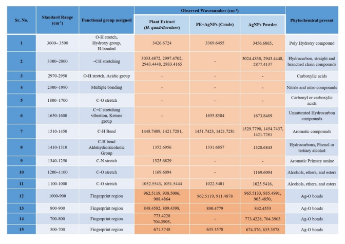
Figure 1: FTIR spectrophotometric analysis of H. quadriloculare extract and synthesized AgNPs
Antifungal Activity of Crude Extracts
Minimum Inhibitory and Fungicidal Concentrations
MIC and MFC assays revealed that the ethanolic and aqueous extracts exhibited lower MIC values than the chloroform and diethyl ether extracts (Figure 2a). Leaf extracts showed the strongest antimicrobial activity, followed by stem and root extracts. MFC values were, in general, higher than MIC values, indicating that crude extracts exerted fungistatic effects predominantly, with complete fungal killing requiring higher concentrations.
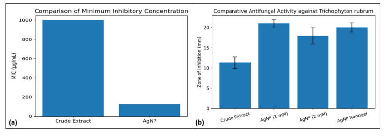
Figure 2(a): Comparison of minimum inhibitory concentration (MIC) of crude extract and silver nanoparticles (AgNPs) against Trichophyton rubrum; (b) Comparative antifungal activity of crude extract, silver nanoparticles, and AgNP-loaded nanogel against Trichophyton rubrum. Values represent mean ± SD (n = 3).
Agar Well Diffusion Assay
Agar well diffusion assays confirmed moderate antifungal activity of crude extracts against Trichophyton rubrum (Figure 2b). Ethanolic stem and leaf extracts produced the largest zones of inhibition (approximately 11-12 mm), while non-polar extracts showed limited activity. The negative control (DMSO) showed no inhibition, confirming assay specificity, while fluconazole exhibited the highest inhibition zone as expected.
Green Synthesis of Silver Nanoparticles
Solar-mediated green synthesis of AgNPs using ethanolic stem extract was successfully achieved, indicated by a rapid colour change from pale yellow to reddish-brown (Figure 3). UV-Vis spectroscopy revealed a characteristic surface plasmon resonance peak at 419.5 nm (Figure 4a), confirming AgNPs formation. Post-purification analysis predicted improved spectral clarity, indicating reduced interference from plant pigments.
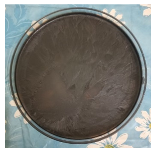
Figure 3: Reddish-brown colored precipitate of AgNPs
Characterization of Silver Nanoparticles
Various detection and characterization techniques were utilized, and physicochemical characterization confirmed successful nanoparticle synthesis as follows. DLS analysis showed Z-average particle sizes between 97-113 nm with moderate polydispersity (PDI ≈ 0.39-0.41) (Figure 4b). Zeta potential values ranged from -11.2 to -11.8 mV, indicating moderate colloidal stability (Figure 4c). SEM images and particle size distribution analysis confirmed predominantly spherical nanoparticles within the 59-112 nm size range (Figure 4d). Particle distribution for synthesized AgNPs is represented as shown in the figure 4e. XRD analysis revealed distinct diffraction peaks corresponding to face-centered cubic (FCC) crystalline silver, with crystallite size estimated at ~18-20 nm (Figure 4f). EDX spectra confirmed the presence of elemental silver, with a characteristic peak near 3 keV.
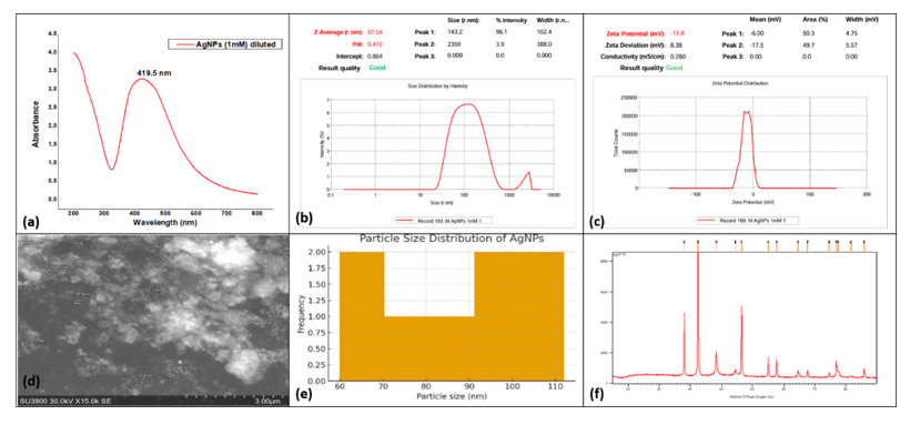
Figure 4 (a): UV-Visible absorption spectrum shows a characteristic surface plasmon resonance (SPR) peak at 419.5 nm; (b) Average zeta size of AgNPs prepared using 1mM AgNO3; (c) Average zeta potential of AgNPs prepared using 1mM AgNO3; (d) FE-SEM micrograph of AgNPs showing particle sizes ranging from ~59 to 112 nm; (e) Particle distribution of AgNPs; (f) X-Ray diffraction pattern of AgNPs indicating FCC crystal structure
Antifungal Activity of Silver Nanoparticles
AgNPs demonstrated significantly greater antifungal activity than crude extracts. The 1 mM AgNPs produced the highest zone of inhibition (21.0 ± 0.9 mm), followed by 2 mM AgNPs (18.0 ± 2.1 mm) (Figure 5a). One-way ANOVA confirmed statistically significant differences among treatment groups (p < 0.01).
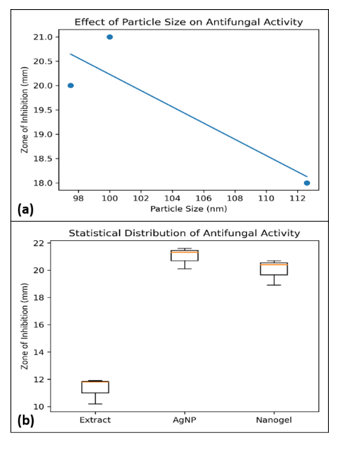
Figure 5(a): Relationship between silver nanoparticle size and antifungal activity, showing increased inhibition with decreasing particle size; (b) Boxplot showing statistical distribution of antifungal activity among crude extract, AgNPs, and AgNP nanogel formulations.
Antifungal Activity of AgNP-Loaded Nanogel
AgNP-loaded nanogel exhibited the strongest antifungal activity. The 1% nanogel formulation produced a zone of inhibition of 20.0 ± 1.1 mm, surpassing AgNPs suspension and crude extract gel. Physicochemical evaluation showed nanogel possessed skin-compatible pH (6.4-6.8), appropriate viscosity (5000-5600 cP), and good spreadability.
Discussion
Dermatophytosis, particularly tinea pedis caused by Trichophyton rubrum, continues to pose a significant therapeutic challenge due to its chronic nature, high recurrence rate, and increasing resistance to conventional antifungal agents. Moreover, antifungal resistance is becoming highly evident in recent times, for instance in case of major cases of dermatophytosis reported from north India. The intensity of infection was displayed in terms of the Dermatophytosis Severity Score (DSS) along with demographic factors and clinical presentations (Lohariwala et al., 2025). The present study demonstrates that the ethno-medicinal plant H. quadriloculare, when integrated with green nanotechnology, exhibits enhanced antifungal efficacy, thereby providing scientific validation to traditional therapeutic practices. This is evident from the recent case studies on T. rubrum infections (Tullio et al., 2025).
Ethnomedicinal relevance and antifungal potential of Heterophragma quadriloculare
The antifungal activity observed for the crude ethanolic and aqueous extracts of H. quadriloculare in the present study is consistent with earlier reports highlighting the antimicrobial potential of members of the Bignoniaceae family. Several species investigations have shown that plants rich in flavonoids, phenolics, tannins, and naphthoquinones exert inhibitory effects on dermatophytes, primarily by interference with fungal cell wall synthesis and membrane integrity (Surana et al., 2014; Mendes de Toledo et al., 2015; Nabatanzi et al., 2020). The predominance of fungistatic activity observed in crude extracts, as reflected by higher MFC values compared to MIC values, aligns with reports on many ethnomedicinal antifungal plants, where therapeutic benefits are often temporary, and relapse is common after treatment discontinuation (Ilkit & Durdu, 2015). Selective antifungal activity is also reported in various extracts derived from leaves making them potential antifungal agents (Kebede & Shibeshi, 2022). The biocatalytic activities are functions of various compounds and secondary metabolites present in the extracts (Gómez-Gaviria et al., 2025).
These findings highlight a critical limitation of traditional herbal therapy: while phytochemicals are biologically active, their low bioavailability, limited penetration into keratinized tissues, and short residence time restrict sustained antifungal action. This underscores the need for formulation-based enhancement rather than dismissal of ethnomedicinal knowledge. The rising incidence of antifungal resistance in dermatophytes, particularly Trichophyton rubrum, further justifies the exploration of alternative nano-enabled therapeutic strategies that operate through multiple mechanisms rather than single molecular targets (Gupta & Versteeg, 2019; Singh & Shukla, 2020).
Enhancement of antifungal efficacy through green-synthesized AgNPs
A key outcome of the present work is the significant enhancement of antifungal activity following green synthesis of AgNPs using H. quadriloculare extract. The observed surface plasmon resonance peak at ~419 nm, spherical morphology, and nanoscale dimensions confirm successful nanoparticle formation, consistent with earlier reports on plant-mediated AgNP synthesis (Shankar et al., 2004; Iravani, 2011). The antifungal potential of green-synthesized AgNPs has been confirmed time to time based on various studies reported in the management of numerous potent fungal pathogens in humans (Huang et al., 2017).
The superior antifungal activity of AgNPs compared to crude extracts observed in this study strongly agrees with multiple investigations demonstrating that nanosilver exhibits broad-spectrum antifungal activity against dermatophytes, including T. rubrum, T. mentagrophytes, and Microsporum species (Kim et al., 2012; Gajbhiye et al., 2009). AgNPs are continuously fabricated and studied for their antifungal potential against various fungal pathogens (Mansoor et al., 2021). Importantly, AgNPs act through multiple mechanisms-membrane disruption, generation of reactive oxygen species, enzyme inactivation, and DNA damage-making resistance development less likely compared to conventional antifungal drugs (Lara et al., 2015; Costa-Orlandi et al., 2021). Being microbiocidal in nature the mechanism of antifungal activity for AgNPs has been approached in a multifaceted manner by various research groups, indicating the sustainable application of AgNPs in various fields (More et al., 2023).
The higher antifungal activity observed with 1 mM AgNPs compared to 2 mM AgNPs in the present study is also supported by previous findings, which show that excessive nanoparticle concentration leads to aggregation, reduced surface area, and diminished biological interactions (Bhattacharjee, 2016). This imposes that antifungal efficacy depends not merely on silver concentration but on optimal particle size, dispersion, and stability (Żarowska et al., 2019).
Phytochemical-nanoparticle synergy: validation of green nanotechnology
The FTIR analysis confirmed the presence of functional groups such as hydroxyl, carbonyl, and amide linkages, indicating that phytochemicals and metabolites from H. quadriloculare served as reducing and capping agents present on the AgNPs. This phytochemical capping plays a critical role in enhancing antifungal activity by improving nanoparticle stability and triggering sustained interaction with fungal cells. Similar synergistic effects have been reported for phytochemical-capped AgNPs synthesized using Azadirachta indica, Ocimum sanctum, and Terminalia species (Ahmed et al., 2016; Mittal et al., 2013; Bamal et al., 2021; Velgosova et al., 2024).
The concept of phytochemical-nanoparticle synergy is precisely important in validating ethnomedicine in the modern context. Rather than bypassing traditional knowledge, green nanotechnology amplifies its efficacy while retaining biocompatibility and environmental safety. This approach supports global efforts to develop sustainable, plant-based nanotherapeutics (WHO, 2019; Karnwal et al., 2024).
Similar enhancements of antimicrobial efficacy through phytochemical-capped silver nanoparticles synthesized using plant extracts have been widely reported, in which plant secondary metabolites act as both reducing and stabilizing agents, resulting in improved biological activity and nanoparticle stability (Ahmed et al., 2016; Mittal et al., 2013). Such green-synthesized nanoparticles are advantageous in antifungal applications due to their biocompatibility and reduced environmental toxicity compared in comparison to chemically synthesized counterparts (Iravani, 2011). Green synthesized AgNPs showed improved antimicrobial properties harbouring selective cytotoxicity, owing to their multifunctional applications (Nayak et al., 2019; Eker et al., 2025).
Improved antifungal performance of AgNP-loaded nanogel formulation
The AgNP-loaded nanogel demonstrated antifungal activity surpassing that of AgNP suspension and crude extract gels, emphasizing the importance of an appropriate drug delivery system. Nanogels enhance topical therapy by improving retention and contact time, ensuring sustained release, and facilitating penetration into infected keratinized tissues. Similar observations have been reported in studies on nanosilver-based topical formulations for fungal infections, in which nanogels outperformed conventional creams and suspensions (Patra et al., 2018; Bala et al., 2020). In account of this, the nanogels are formulated as an efficient drug delivery platform specifically for dermal and transdermal delivery (Siafaka et al., 2023). Being an important active medicinal constituent AgNPs are utilized as a multifunctional entities in wound healing directly or as a part of wound dressings and ointments (Balaure et al., 2025).
The skin-compatible pH and the nanogel's observed rheological properties further support its potential for topical application. The statistically significant improvement in antifungal activity (p < 0.05) confirms that the formulation strategy contributed meaningfully to therapeutic enhancement rather than serving as a passive carrier.
Statistical validation and clinical relevance
Statistical analysis based upon one-way ANOVA and independent t-tests confirmed that differences among crude extracts, AgNPs, and nanogel formulations were significant and not due to random variation. The observed negative correlation between nanoparticle size and antifungal activity is consistent with established nanotoxicological principles, in which a reduction in particle size increases surface area and biological interactions and catalysis potential (Elechiguerra et al., 2005).
From a clinical perspective, these findings are highly relevant. Chronic and recurrent tinea pedis is increasingly reported in India and other tropical regions, often requiring prolonged antifungal therapy with limited success (Verma & Madhu, 2017). The nano-enabled ethnomedicinal approach presented here offers a promising alternative that could minimize treatment duration, recurrence, and dependence on synthetic antifungals.
One-way ANOVA predicted a statistically significant difference (p < 0.05) in antifungal activity among crude plant extracts, green-synthesized AgNPs, and AgNP-loaded nanogel formulations against Trichophyton rubrum. The mean zone of inhibition produced by crude ethanolic extracts was significantly lower than that observed for AgNPs, confirming the limited fungistatic potential of crude extracts.
Pairwise t-test analysis showed that AgNPs (21.0 ± 0.9 mm) exhibited significantly higher antifungal activity than crude extracts (p < 0.01). Furthermore, the AgNP-loaded nanogel (20.0 ± 1.1 mm) demonstrated a statistically comparable antifungal effect to AgNP suspension (p > 0.05), indicating that incorporation into the nanogel matrix did not reduce biological efficacy while improving formulation stability and applicability.
The observed variation between 1 mM and 2 mM AgNP concentrations was also statistically significant (p < 0.05), with the 1 mM formulation showing superior antifungal activity. This suggests that optimal nanoparticle dispersion and surface availability, rather than higher silver concentration alone, plays a crucial role in antifungal efficacy.
Overall, the statistical analysis confirms the enhancement in antifungal activity achieved through green synthesis of AgNPs and subsequent nanogel formulation is significant and reproducible, thereby validating the effectiveness of the nano-enabled ethnomedicinal approach.
Need for validating ethnomedicine through green nanotechnology
In the current era of antimicrobial resistance, environmental concerns, and rising healthcare costs, validating ethnomedicine through green nanotechnology is not merely innovative but necessary. Traditional remedies, when scientifically optimized and standardized through nanotechnology, can provide affordable, effective, and culturally acceptable therapeutic solutions. The present study exemplifies this integration by transforming a traditionally fungistatic plant remedy into a potent nano-enabled antifungal formulation. This suggests applicability of green-synthesized AgNPs as a sustainable strategy for the management of antimicrobial resistance (Törős et al., 2025). Thus, green nanotechnology is gaining rigorous attention in healthcare applications, advancing the innovative applications and use of eco-friendly nanoparticles like biogenic AgNPs (Ayub et al., 2025).
Conclusion
This study demonstrates that Heterophragma quadriloculare exhibits antifungal activity against Trichophyton rubrum, which is significantly enhanced through green synthesis of AgNPs. While crude plant extracts showed moderate, mainly fungistatic effects, phytochemical-capped AgNPs displayed superior antifungal efficacy. Physicochemical characterization confirmed the formation of stable nanoparticles, and statistical analysis validated the significance of the observed improvements. The AgNP-loaded nanogel further enhanced antifungal activity and exhibited suitable properties for topical application. Overall, the findings provide scientific validation of ethnomedicinal knowledge and highlight green nanotechnology as an effective strategy to improve traditional antifungal therapies. Further in vivo and clinical studies are required to confirm therapeutic applicability.
Acknowledgement
The authors gratefully acknowledge the Rajiv Gandhi Science and Technology Commission (RGSTC), Mumbai, Maharashtra and University of Mumbai for providing financial support under the scheme of Assistance for Science and Technology Applications and for granting an extension to complete the research work.
References
- Agrawal S, Singh NK, Neupane S, et al NEXTODERM: Consensus on Dermatophytosis Diagnosis and Management in Nepal. Dermat Res Pract(2025): 1347872.
- Ahmed S, Ahmad M, Swami BL, et al. A review on plants extract mediated synthesis of silver nanoparticles for antimicrobial applications: A green expertise. J Adv Res 7 (2016): 17-28.
- Ameen M. Epidemiology of superficial fungal infections. Clinics Dermato 28 (2010): 197-201.
- Amenu D, Desalegn R, Nugusa A, et al. Evaluation of antifungal activity of plant extracts against plant pathogen associated with sweet orange (Citrus sinensisL) in Jimma Western Ethiopia Dis Appl Sci 6 (2024): 357.
- Ayatollahi Mousavi SA, Salari S, Hadizadeh S. Evaluation of Antifungal Effect of Silver Nanoparticles Against Microsporum canis, Trichophyton mentagrophytesand Microsporum gypseum. Iran J Biotech 13 (2015): 38–42.
- Ayub A, Wani AK, Malik SM, et al. Green nanoscience for healthcare: Advancing biomedical innovation through eco-synthesized nanoparticle. Biotech Rep (Amsterdam, Netherlands)47 (2025): e00913.
- Balaure PC, Niculescu AG, Anghel D, et al Role of Silver Nanoparticles in Wound Healing: Mechanisms, Efficacy, and Clinical Applications. Inorganics13 (2025): 401.
- Bamal D, Singh A, Chaudhary G, et al Silver Nanoparticles Biosynthesis, Characterization, Antimicrobial Activities, Applications, Cytotoxicity and Safety Issues: An Updated Review. Nanomaterials (Basel, Switzerland)11 (2021): 2086.
- Bhattacharjee S DLS and zeta potential - What they are and what they are not? J Control Rel 235 (2016): 337-351.
- Clinical and Laboratory Standards Institute Reference method for broth dilution antifungal susceptibility testing of filamentous fungi (CLSI document M38-A2). CLSI, Wayne, PA (2008).
- Costa-Orlandi CB, Sardi JCO, Santos CT, et al Fungal biofilms and polymicrobial diseases. J Fungi 7 (2021): 201.
- Cowan MM Plant products as antimicrobial agents. Clin Micro Rev 12 (1999): 564-582.
- Eker F, Akdaşçi E, Duman H, et al. Green Synthesis of Silver Nanoparticles Using Plant Extracts: A Comprehensive Review of Physicochemical Properties and Multifunctional Applications. Int J Mol Sci26 (2025): 6222.
- Elechiguerra JL, Burt JL, Morones JR, et al. Interaction of silver nanoparticles with HIV-1. J Nanobiotech 3 (2005): 6.
- Gajbhiye M, Kesharwani J, Ingle A, et al. Fungicidal activity of silver nanoparticles synthesized by aqueous leaf extract of Ocimum sanctum against phytopathogenic fungi. Nanomed Nanotech Biol Med 5 (2009): 382-386.
- Gómez-Gaviria M, Baruch-Martínez DA, Mora-Montes HM. Natural Source-Derived Compounds with Antifungal Activity Against Medically Relevant Fungi.Infec Drug Resist 18 (2025): 6389–6406.
- Gupta AK, Versteeg SG. Topical treatment of superficial fungal infections. Drugs 79 (2019): 367-380.
- Gutiérrez-Pinzón Y, Martínez-Preciado AH, Velázquez-López JM, et al. Antimicrobial Potential of Nanomaterials Synthesized with Extracts from AnnonaPlants: A Review. Antibiotics (Basel, Switzerland) 14 (2025): 748.
- Harborne JB. Phytochemical methods: A guide to modern techniques of plant analysis (3rd ed) Springer (1998).
- Havlickova B, Czaika VA, Friedrich M Epidemiological trends in skin mycoses worldwide. Mycoses 51 (2008): 2-15.
- Hernández-Ceja A, Loeza-Lara PD, Espinosa-García FJ, et al. In Vitro Antifungal Activity of Plant Extracts on Pathogenic Fungi of Blueberry (Vaccinium). Plants (Basel, Switzerland)10 (2021): 852.
- Huang W, Bao Y, Duan H, et al Antifungal effect of green synthesised silver nanoparticles against Setosphaeria turcica. IET Nanobiotech11 (2017): 803–808.
- Ilkit M, Durdu M Tinea pedis: The etiology and global epidemiology. J Fungi 1 (2015): 9-21.
- Iravani S. Green synthesis of metal nanoparticles using plants. Green Chem 13 (2011): 2638-2650.
- Jangid H, Singh S, Kashyap P, et al. Advancing biomedical applications: an in-depth analysis of silver nanoparticles in antimicrobial, anticancer, and wound healing roles. Front Pharmacol15 (2024): 1438227.
- Jangjou A, Zareshahrabadi Z, Abbasi M, et al. Time to Conquer Fungal Infectious Diseases: Employing Nanoparticles as Powerful and Versatile Antifungal Nanosystems against a Wide Variety of Fungal Species. Sustainability14 (2022): 12942.
- Karnwal A, Jassim AY, Mohammed AA, et al. Nanotechnology for Healthcare: Plant-Derived Nanoparticles in Disease Treatment and Regenerative Medicine. Pharmaceuticals (Basel, Switzerland)17 (2024): 1711.
- Kebede B, Shibeshi W. In vitro antibacterial and antifungal activities of extracts and fractions of leaves of Ricinus communis Linn against selected pathogens. Vet Med Sci8 (2022): 1802–1815.
- Kim SW, Jung JH, Lamsal K, et al. Antifungal effects of silver nanoparticles against dermatophytes. Mycobiology 40 (2012): 53-58.
- Kokate CK. Practical pharmacognosy (4th ed.). Vallabh Prakashan (2005).
- Lara HH, Ayala-Núñez NV, Ixtepan-Turrent L, et al. Silver nanoparticles as antimicrobial agents: A review. World J Micro Biotech 31 (2015): 1821-1832.
- Leung AK, Barankin B, Lam JM, et al. Tinea pedis: an updated review. Drugs Cont12 (2023): 2023-5-1.
- Lohariwala A, Gupta S, Kaur N, et al. Emerging Antifungal Resistance in Dermatophytosis: A Clinicomycological Study from North India. Cureus17 (2025): e92679.
- Mansoor S, Zahoor I, Baba TR, et al. Fabrication of Silver Nanoparticles Against Fungal Pathogens. Front Nanotechnol3 (2021): 679358.
- Mendes de Toledo CE, Santos PR, Palazzo de Mello JC, et al. Antifungal Properties of Crude Extracts, Fractions, and Purified Compounds from Bark of Curatella americana (Dilleniaceae) against Candida Species. Evidence-based Compl Alter Med eCAM (2015): 673962.
- Mittal AK, Chisti Y, Banerjee UC. Synthesis of metallic nanoparticles using plant extracts. Biotech Adv 31 (2013): 346-356.
- More PR, Pandit S, Filippis A, et al. Silver Nanoparticles: Bactericidal and Mechanistic Approach against Drug Resistant Pathogens. Microorganisms11 (2023): 369.
- Nabatanzi A, Nkadimeng MS, Lall N, et al. Ethnobotany, Phytochemistry and Pharmacological Activity of Kigelia africana(Lam.) Benth. (Bignoniaceae). Plants (Basel, Switzerland) 9 (2020): 753.
- Nayak PS, Pradhan S, Arakha M, et al. Silver nanoparticles fabricated using medicinal plant extracts show enhanced antimicrobial and selective cytotoxic propensities. IET Nanobiotech13 (2019): 193–201.
- Patra JK, Das G, Fraceto LF, et al Nano-based drug delivery systems: Recent developments and future prospects. J Nanobiotech 16 (2018): 71.
- Pfaller MA, Diekema DJ, Andes D, et al. Clinical breakpoints for antifungal susceptibility testing. Clin Micro Rev 23 (2010): 160-179.
- Policepatel SS, Manikrao VG. Ethnomedicinal plants used in the treatment of skin diseases in Hyderabad Karnataka region, Karnataka, India. Asian Pacific J Trop Biomed3 (2013): 882–886.
- Rodrigues AS, Batista JGS, Rodrigues MÁV, et al. Advances in silver nanoparticles: a comprehensive review on their potential as antimicrobial agents and their mechanisms of action elucidated by proteomics. Front Microbiol 15 (2024): 1440065.
- Sable PD, Hossain E, Suple SD, et al. Traditional treatments for skin ailments: a review of ethnomedicinal plants used to treat skin infections. Zenodo (2025).
- Satani NM, Thite AG, Kore KJ. Ethnomedicinal plants used for skin diseases in Maharashtra. Int J Herb Med 4 (2016): 45-52.
- Shankar SS, Rai A, Ahmad A, et al. Rapid synthesis of Au, Ag, and bimetallic nanoparticles using neem (Azadirachta indica) leaf broth. J Coll Interface Sci 275 (2004): 496-502.
- Siafaka PI, Özcan Bülbül E, Okur ME, et al. The Application of Nanogels as Efficient Drug Delivery Platforms for Dermal/Transdermal Delivery. Gels (Basel, Switzerland)9 (2023): 753.
- Singh A, Shukla P Antifungal resistance in dermatophytes: Mechanisms and implications. Mycoses 63 (2020): 1028-1035.
- Singh J, Kumar A, Nayal AS, et al. Comprehensive antifungal investigation of green synthesized silver nanoformulation against four agriculturally significant fungi and its cytotoxic applications. Sci Rep14 (2024): 5934.
- Sousa F, Ferreira D, Reis S, et al. Current Insights on Antifungal Therapy: Novel Nanotechnology Approaches for Drug Delivery Systems and New Drugs from Natural Sources. Pharmaceuticals (Basel, Switzerland)13 (2020): 248.
- Summerbell RC, Kane J, Krajden S Onychomycosis, tinea pedis, and tinea manuum caused by dermatophytes. Clin Micro Rev 18 (2005): 73-101.
- Surana SJ, Gokhale SB, Jadhav RB Phytochemical and antimicrobial evaluation of Heterophragma quadriloculare. Pharmacognosy J 6 (2014): 12-18.
- Thatyana M, Dube NP, Kemboi D, et al. Advances in Phytonanotechnology: A Plant-Mediated Green Synthesis of Metal Nanoparticles Using PhyllanthusPlant Extracts and Their Antimicrobial and Anticancer Applications. Nanomaterials 13 (2023): 2616.
- Todorova M, Petrova V, Ranguelov B, et al. Green Synthesis of Antimicrobial Silver Nanoparticles (AgNPs) from the Mucus of the Garden Snail Cornu aspersum. Molecules (Basel, Switzerland)30 (2025): 2150.
- Törős G, El-Ramady H, Nguyen DHH, et al. Green-Synthesized Nanomaterials from Edible and Medicinal Mushrooms: A Sustainable Strategy Against Antimicrobial Resistance. Pharmaceutics17 (2025): 1388.
- Tullio V, Panzone M, Cervetti O, et al. Case Report: From Misdiagnosis to Accurate Identification: Managing a Case Series of Trichophyton rubrumMicroorganisms 13 (2025): 895.
- Velgosova O, Dolinská S, Podolská H, et al. Impact of Plant Extract Phytochemicals on the Synthesis of Silver Nanoparticles. Materials17 (2024): 2252.
- Verma S, Madhu R. The great Indian epidemic of superficial dermatophytosis. Ind J Dermat 62 (2017): 227-236.
- Ward H, Parkes N, Smith C, et al. Consensus for the Treatment of Tinea Pedis: A Systematic Review of Randomised Controlled Trials. J Fungi8 (2022): 351.
- World Health Organization WHO guidelines on good agricultural and collection practices (GACP) for medicinal plants. WHO Press (2003).
- World Health Organization. WHO global report on traditional and complementary medicine 2019. Geneva: WHO (2019).
- Żarowska B, Koźlecki T, Piegza M, et al. New Look on Antifungal Activity of Silver Nanoparticles (AgNPs). Polish J Micro68 (2019): 515–525.