Maryam Naser1, Farbod Nasseri2, Nafiseh Hashemi1*
1Hashemi Eye Care, Encino, CA, USA.
2Renaissance Imagine Medical Associates, Northridge, CA, USA.
*Corresponding Author: Nafiseh Hashemi, MD, Hashemi Eye Care, 5353 Balboa Blvd #110, Encino, CA, USA
Received: 03 September 2020; Accepted: 14 September 2020; Published: 18 September 2020
DOI: 10.26502/fjor.2644-00240021
ShareIntroduction
Pituitary adenomas are the most common cause of optic chiasm compression in adults. These tumors are classified by their size to Microadenomas: <10 mm and Macroadenomas: ≥10 mm which is diagnosed by CT or MRI. Peripheral vision loss, blurry vision, diplopia, headache and dizziness are the usual symptoms of pituitary tumor. We are presenting a patient with peripheral vision loss and optic nerve damage from cystic compression of pituitary macroadenoma.
Cystic compression; Pituitary Adenoma; Peripheral vision loss; Blurry Vision; Bitemporal visual field defect
Patient and Observation
The patient is a 45 year-old male who is referred to neuro-ophthalmology clinic complaining of having blurred vision on both eyes. He noticed bilateral progressive peripheral vision loss started in the left eye since 4 months ago. He denies headaches, diplopia, head trauma, infectious or inflammatory diseases. He does not have any past medical or surgery history. He is not using any medication.
Vision is 20/25 right eye and 20/30 left eye. Intra ocular pressure is 13 mmHg. Color vision is 11/11 right eye and 8/11 left eye. Visual field shows bitemoporal field defect (Figure 1). OCT optic nerve presents normal RNFL thickness, even though the patient has RAPD and color vision deficit left eye (Figure 2). The funds photo in left eye shows temporal atrophy (Figure 3). Since the presence of bitemporal visual field defect is pathognomonic of chiasmal compression, MRI brain was ordered which revealed cystic macroadenoma 3.3cm x 3.3cm x 3.4cm with compression over chiasma (Figure 4). The patient was referred to neurosurgeon for further evaluation and treatment.
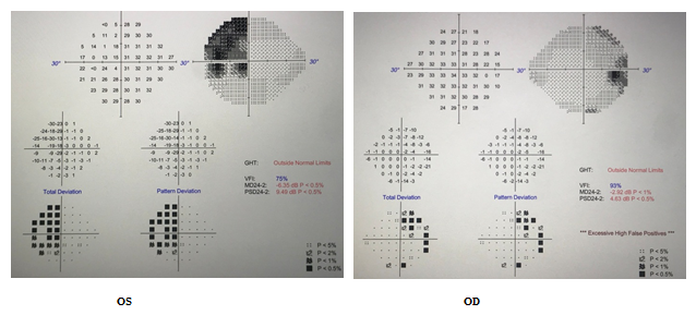
Figure1: VF bitemporal field defect
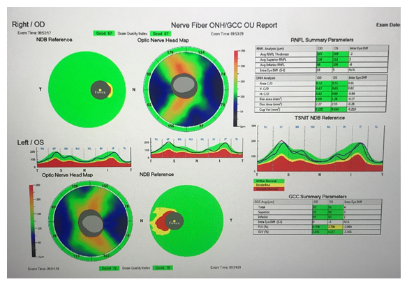
Figure 2: OCT Optic Nerve: Normal average RNFL thickness, mild GCL loss OS
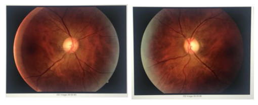
Figure 3: Fundus Photos: mild temporal atrophy left eye
Figure 4: There is 3.3cm x 3.3cm x 3.4cm (AP x transverse x craniocaudal) sellar/suprasellar predominantly cystic mass with associated fluid-fluid level most consistent with a cystic macroadenoma. There is significant mass effect on optic chiasm.
Discussion
Pituitary adenomas are usually benign, slow-growing tumors that arise from cells in the pituitary gland and count about 16% of all primary cranial neoplasms [1,8]. Study on 33cases with pituitary macroadenoma showed almost equal sex incidence with average age group of 50 to 60 years old. The common complaints were headache and vision loss [2]. The patients may experience different signs and symptoms based on mass effect of the tumor versus hormone secretion from functioning pituitary adenoma [3]. In our case the patient have not had any headaches and the only complaint was difficulty changing lane when he was driving. Imaging plays an important role in diagnosis of sellar lesions such as pituitary tumors [4]. Long-term compression of the optic chiasma may lead to axonal degeneration of ganglion cells. Spectral domain optical coherence tomography (SD-OCT) is useful to verify retinal nerve fiber layer and ganglion cell loss in the retina. OCT may even help in diagnose of asymptomatic pituitary macroadenoma [5]. In our case, OCT did not show RNFL thinning but GCL loss on the left retina. The treatment of macro adenoma is based on the type of the tumor. In non-secretory macroadenomas, surgical extirpation is often required. Trans-sphenoidal surgery is preferred approach [6,7]. Functioning pituitary adenomas with variant clinical syndromes need on-time treatment to reduce associated morbidity and mortality. Treatment approaches include trans-sphenoidal surgery, medical therapy and radiation [8,9,10].
Conclusion
Predominantly cystic macroadenomas may cause peripheral vision loss and optic nerve damage with relatively normal looking optic nerve and normal OCT which may delay the diagnosis. Management of pituitary adenoma can vary based on the type of tumor and require cooperation of ophthalmologist, radiologist, endocrinologist, neurosurgeon and pathologist.
References
- Sharif-Alhoseini M, Rahimi-Movaghar V. Pituitary adenomas: a review. Journal of Injury and Violence Research 4 (2012).
- Gupta K, Sahni S, Saggar K, et al. Evaluation of clinical and magnetic resonance imaging profile of pituitary macroadenoma: A prospective study. Journal of Natural Science, Biology, and Medicine 9 (2018): 34.
- Lim CT, Korbonits M. Update on the clinicopathology of pituitary adenomas. Endocrine Practice 24 (2018): 473-488.
- Karimian-Jazi K. Pituitary gland tumors. Der Radiologe (2019).
- Asensio-Sánchez VM, Foncubierta J. Progressive loss of vision caused by asymptomatic pituitary macroadenoma: role of OCT. International Medical Case Reports Journal 9 (2016): 291.
- Wu JS, Shou XF, Yao CJ, et al. Transsphenoidal pituitary macroadenomas resection guided by PoleStar N20 low-field intraoperative magnetic resonance imaging: comparison with early postoperative high-field magnetic resonance imaging. Neurosurgery 65 (2009): 63-71.
- Theodosopoulos PV, Leach J, Kerr RG, et al. Maximizing the extent of tumor resection during transsphenoidal surgery for pituitary macroadenomas: can endoscopy replace intraoperative magnetic resonance imaging?. Journal of Neurosurgery 112 (2010): 736-743.
- Katznelson L, Laws Jr ER, Melmed S, et al. Acromegaly: an endocrine society clinical practice guideline. The Journal of Clinical Endocrinology & Metabolism 99 (2014): 3933-3951.
- Nieman LK, Biller BM, Findling JW, et al. Treatment of Cushing's syndrome: an endocrine society clinical practice guideline. The Journal of Clinical Endocrinology & Metabolism 100 (2015): 2807-2831.
- Varlamov EV, McCartney S, Fleseriu M. Functioning pituitary adenomas–current treatment options and emerging medical therapies. European Endocrinology 15 (2019): 30.