Suvarthy Dey1, Sabrina Sifat2, Farjana Sharmeen Shimu3, Koli Das4, Maisha Farzana Rahman5, Farah Habibur Rahman6, Aysha Siddika Rina7, Abdullah Al Masud1, Avijit Sarker Jyoti1,5*
1Dream USMLE
2Kumudini Women's Medical College and Hospital, Bangladesh
3National Institute of Ophthalmology and Hospital, Dhaka, Bangladesh
4Chittagong medical college Hospital, Bangladesh
5Mugda Medical College, Dhaka, Bangladesh
6Zainul Haque Sikder women’s medical college and Hospital, Dhaka, Bangladesh
7Dinajpur Medical College, Dinajpur, Bangladesh
*Corresponding Author: Avijit Sarker Jyoti, Dream USMLE, Mugda Medical College, Dhaka, Bangladesh.
Received: 06 July 2026; Accepted: 12 August 2026; Published: 11 September 2026.
Background: Congenital chikungunya is an uncommon consequence of vertical transmission of chikungunya virus (CHIKV), typically occurring when maternal infection develops during the intrapartum period. Because affected neonates often present with nonspecific manifestations such as respiratory distress, poor perfusion, and elevated inflammatory markers, the condition may closely resemble early-onset neonatal sepsis. Characteristic cutaneous hyperpigmentation, although increasingly recognized, remains an underreported clinical sign that may facilitate early diagnosis.
Case Presentation: We report a full-term male neonate born by lower uterine cesarean section to a 29-year-old mother who developed RT-PCR-confirmed chikungunya infection with high-grade fever and generalized rash one day before delivery. Soon after birth, the neonate developed progressive respiratory distress requiring admission to the neonatal intensive care unit. Initial evaluation favored early-onset neonatal sepsis because of respiratory compromise and markedly elevated serum procalcitonin despite negative blood cultures. The infant received empirical broad-spectrum antibiotics and supportive management. During hospitalization, respiratory distress gradually resolved; however, the neonate subsequently developed diffuse blackish cutaneous hyperpigmentation associated with neonatal jaundice. Given the maternal history and ongoing regional chikungunya activity, congenital CHIKV infection was suspected and confirmed by neonatal RT-PCR. Antibiotics were discontinued after exclusion of bacterial infection, and the infant was managed conservatively with supportive care, resulting in complete clinical recovery and gradual resolution of skin pigmentation. He was discharged in satisfactory condition with advice for long-term neurodevelopmental follow-up.
Conclusion: This case highlights generalized blackish hyperpigmentation as an important clinical clue to congenital chikungunya in neonates presenting with sepsis-like illness. Recognition of this distinctive cutaneous manifestation, together with careful maternal history and timely virological testing, may facilitate early diagnosis, reduce unnecessary antibiotic exposure, and improve clinical management, particularly in regions where chikungunya is endemic or epidemic.
Congenital chikungunya; Neonatal hyperpigmentation; Early-onset neonatal sepsis; Vertical transmission
Congenital chikungunya articles; Neonatal hyperpigmentation articles; Early-onset neonatal sepsis articles; Vertical transmission articles.
1. Introduction
Chikungunya is a mosquito-borne viral disease caused by the chikungunya virus (CHIKV), an enveloped, positive-sense single-stranded RNA virus belonging to the genus Alphavirus of the family Togaviridae. Transmitted primarily by Aedes aegypti and Aedes albopictus, CHIKV has re-emerged as an important public health problem in tropical and subtropical regions, including South Asia, following repeated epidemics over the past two decades [1,2]. Although chikungunya is generally a self-limiting illness characterized by fever, arthralgia, myalgia, and rash in adults, maternal infection during late pregnancy carries a substantial risk of vertical transmission, particularly when maternal viremia coincides with delivery [2-4].
Congenital chikungunya is an uncommon but increasingly recognized neonatal infection. Vertical transmission occurs predominantly during the intrapartum period, with nearly half of neonates born to viremic mothers at delivery becoming infected [3,4]. Affected infants are usually asymptomatic at birth but develop clinical manifestations between the third and seventh day of life, including fever, respiratory distress, poor feeding, apnea, thrombocytopenia, myocarditis, encephalopathy, seizures, shock, and multiorgan dysfunction [3-5]. These manifestations frequently overlap with those of early-onset neonatal sepsis, posing a significant diagnostic challenge, particularly in resource limited settings where bacterial sepsis is more prevalent and viral diagnostics are not routinely performed [3,5,6]. Among the diverse manifestations of congenital chikungunya, cutaneous hyperpigmentation is considered one of the most distinctive yet underrecognized clinical signs. Classically described as the "Chik sign" or "brownie nose," hyperpigmentation usually appears several days after birth and may involve the nose, face, trunk, and extremities [7,8]. Although the exact pathophysiological mechanism remains unclear, inflammatory injury of the dermoepidermal junction with pigmentary incontinence and cytokine-mediated melanocyte stimulation has been proposed [7,8]. Recognition of this characteristic dermatological finding may provide an invaluable clinical clue for distinguishing congenital chikungunya from neonatal bacterial sepsis, particularly during outbreaks or in neonates born to mothers with recent febrile exanthematous illness.
Bangladesh has experienced recurrent chikungunya outbreaks in recent years, increasing the risk of maternal and neonatal infections [9]. Nevertheless, reports of RT-PCR-confirmed congenital chikungunya presenting with diffuse generalized blackish hyperpigmentation remain scarce. Furthermore, generalized hyperpigmentation accompanying respiratory distress and elevated inflammatory biomarkers can closely mimic early-onset neonatal sepsis, potentially leading to delayed diagnosis and unnecessary prolonged antibiotic therapy. We report a neonate with RT-PCR-confirmed congenital chikungunya who initially presented with severe respiratory distress and sepsis-like illness but subsequently developed progressive generalized blackish hyperpigmentation, highlighting this distinctive cutaneous manifestation as an important diagnostic clue. This case emphasizes the importance of obtaining a detailed maternal history, considering congenital chikungunya in the differential diagnosis of neonatal sepsis during outbreaks, and recognizing characteristic skin pigmentation as an early clinical marker that may facilitate timely diagnosis and appropriate supportive management.
2. Case Presentation
A 50-hour old male neonate, first issue of a non-consanguineous couple, delivered at 39 weeks of gestation via lower uterine caesarean section (LUCS) got admitted into “X” hospital with the complaints off respiratory distress since birth and blackish coloration of his skin from 3rd day of his age to onwards. His mother was 29 years old with no bad obstetrical history and had an irregular anti-natal check-up but immunized against tetanus and as per EPI schedule. The pregnancy was complicated by her pre-existing hypothyroidism but there was no history of gestational hypertension, gestational diabetes mellitus or pre-mature rupture of membrane. Her pregnancy was uneventful up to 38 weeks of gestation. During her current pregnancy, she reported a febrile illness with a rash all over body approximately one day before delivery. The fever continued, high grade in nature, highest recorded temperature was 104◦F, subsided after taking antipyretics, associated with chills and rigor, myalgia, body-ache and malaise and rashes all over body. The rashes appeared in hands and feet, and trunk first then subsequently spread over various parts of body but spared the face. She was tested negative for Dengue by ELISA method but positive for chikungunya virus by RT-PCR method. The pregnancy was complicated by oligohydramnios and with bipedal pitting oedema from the 25th week of pregnancy. At her 38 weeks of gestation, she delivered a male baby weighing 3,000 g, which cried immediately after the birth but developed progressive respiratory distress evidenced by repeated shallow breathing, chest indrawing, grunting Immediately, as per resuscitation protocol, the baby was managed appropriately and treated with high flow oxygen but the condition didn’t improve. So, the baby was transferred to neonatal intensive care unit and examined. During examination, the baby was pink in appearance with good activity and reflexes, there was peripheral cyanosis, cold periphery, capillary refill time-4 sec, heart rate-140 bpm, respiratory rate- 65 breaths per minute, with shallow breathing pattern, air entry was equal on both lungs, both first and second heart sound was noted, abdomen was soft, umbilicus was healthy, capillary blood glucose-3.7 mmol/L, OFC was 33 cm, length-45 cm. Initial clinical assessment favored a diagnosis of neonatal sepsis. Appropriate investigations including blood culture were sent to laboratory and empiric intravenous antibiotics, supportive management were initiated by NICU protocol. Laboratory investigations revealed the following hematological parameters: Hemoglobin: 16.3 g/dL; total leukocyte count: 14.74 × 10⁹/L, neutrophils: 58.4%; lymphocytes: 30.1%; platelet count: 270 × 10⁹/L; hematocrit: 46.9%. [Table 1] Echocardiography revealed a very tiny outlet ventricular septal defect (VSD) with a left-to-right shunt and a patent foramen ovale (PFO) with bi-directional shunting and good biventricular function. Peripheral blood film examination revealed no abnormality, but biochemical investigations showed markedly elevated procalcitonin levels (8.80 ng/mL without any growth in blood culture. The neonate was initially managed empirically for suspected neonatal sepsis with intravenous antibiotics (Ceftazidime, Amikacin, and Levofloxacin). The respiratory distress resolved gradually but the baby developed neonatal jaundice evidenced by yellowish coloration of skin and eyes. So, he was treated with hydration and nutritional support along with phototherapy. After 9 days of his age, the jaundice resolved but there was blackish pigmented skin lesion over his body including Chik sign (Figure1). Considering maternal febrile illness and the ongoing chikungunya activity in the region, congenital chikungunya infection was suspected and there was positive RT PCR for CHIKV.
Following the confirmation of CHIKV and negative bacterial cultures, management shifted to supportive care. The neonate was managed conservatively with supportive care, including fluid management, temperature monitoring, and nutritional support. The infant showed progressive clinical improvement, with resolution of respiratory distress and stabilization of vital parameters and skin changes to normal gradually. He was discharged after 16 days of inpatient care in satisfactory condition. Long-term neurodevelopmental follow-up was advised given the risk of subclinical injury associated with neonatal CHIKV infection.
Table 1: Investigation profile of the neonate.
|
Investigation Profile |
|
|
Hemoglobin |
16.30% |
|
Total Leukocytes count |
14.74 × 10⁹/L |
|
Neutrophils |
58.40% |
|
Lymphocytes |
30.10% |
|
Platelet count |
270 × 10⁹/L |
|
Hematocrit |
46.90% |
|
Inflammatory |
|
|
Procalcitonin |
8.80 ng/ml (normal <0.50 ng/ml) |
|
Serum Electrolytes |
|
|
sodium |
149 mmol/L |
|
calcium |
2.3 mmol/L |
|
Biochemistry |
|
|
Serum creatinine |
75 µmol/L |
|
Blood culture |
No growth after five days of incubation |
|
Virology (RT-PCR) |
|
|
Chikungunya Virus RNA |
Positive |
|
Dengue |
Negative |
|
Zika virus |
Negative |
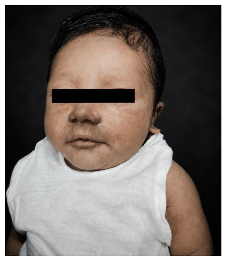
Figure 1: Blacking hyperpigmentation of the neonate (Chick sign).
3. Discussion
Congenital chikungunya is an uncommon but increasingly recognized consequence of vertical transmission of chikungunya virus (CHIKV), particularly when maternal infection occurs during the intrapartum period while maternal viremia is present [3,4]. Vertical transmission is believed to occur trans-placentally around the time of delivery, and affected neonates usually become symptomatic within the first week of life. Typical manifestations include fever, respiratory distress, poor feeding, irritability, thrombocytopenia, hemodynamic instability, neurological involvement, and characteristic cutaneous manifestations [3,5]. In the present case, the mother developed high-grade fever with generalized rash one day before delivery and was confirmed to have acute CHIKV infection by RT-PCR. Subsequently, the neonate developed respiratory distress soon after birth followed by progressive blackish cutaneous hyperpigmentation, while neonatal RT-PCR also confirmed CHIKV infection, strongly supporting congenital transmission.
An important and unique feature of this case was the development of progressive generalized blackish hyperpigmentation. Hyperpigmentation is increasingly recognized as a characteristic dermatological manifestation of neonatal chikungunya and has been described as the "Chik sign" or "brownie nose," although diffuse pigmentation involving the trunk and extremities has also been reported [7,8]. The exact pathogenesis remains uncertain. Proposed mechanisms include virus-induced inflammation at the dermo-epidermal junction, melanocyte stimulation by inflammatory cytokines, post-inflammatory melanosis, and pigmentary incontinence resulting from epidermal injury [7,8]. Unlike bacterial sepsis, where diffuse hyperpigmentation is uncommon, this distinctive pigmentation can provide an important clinical clue toward congenital chikungunya, particularly in endemic regions experiencing outbreaks. In our patient, generalized blackish pigmentation developed after improvement of the acute respiratory illness and gradually resolved with supportive care, a clinical course consistent with previously published reports [8].
The initial presentation closely resembled early-onset neonatal sepsis. Respiratory distress, prolonged capillary refill time, peripheral cyanosis, and elevated inflammatory biomarkers prompted empirical broad-spectrum antibiotic therapy according to standard neonatal practice. Such diagnostic confusion has been frequently reported because congenital chikungunya shares several clinical features with bacterial sepsis during the early neonatal period (3, 5). However, several findings in our case favored congenital CHIKV infection over bacterial sepsis. First, there was a clear epidemiological link with laboratory-confirmed maternal chikungunya immediately before delivery. Second, blood cultures remained sterile despite significant clinical illness. Third, hematological findings did not demonstrate marked neutrophilic leukocytosis or persistent thrombocytopenia that would strongly support invasive bacterial infection. Finally, the appearance of progressive generalized hyperpigmentation together with positive neonatal RT-PCR for CHIKV established the diagnosis. These observations emphasize that congenital chikungunya should be considered neonates with sepsis-like illness born to mothers with peripartum febrile rash illness, especially during outbreaks [3,4].
Respiratory distress is one of the recognized manifestations of congenital chikungunya and has been described in several neonatal case series [3,5]. Although its exact mechanism is incompletely understood, it is likely multifactorial. Systemic inflammatory response induced by viral replication may increase pulmonary vascular permeability, resulting in transient pulmonary dysfunction, while myocardial involvement, capillary leak, endothelial dysfunction, and generalized inflammation may further contribute to respiratory compromise [3]. In severe diseases, cardiovascular instability and multiorgan involvement may also aggravate respiratory symptoms. Fortunately, most affected neonates improve with supportive management without requiring disease-specific antiviral therapy, as observed in our patient [4]. Another interesting finding in this case was markedly elevated serum procalcitonin despite sterile blood cultures. Although procalcitonin is widely used as a biomarker of bacterial sepsis, elevated concentrations are not entirely specific for bacterial infection. Viral infections associated with significant systemic inflammation, tissue injury, or severe respiratory disorders may also produce elevated procalcitonin levels [10]. Furthermore, neonatal respiratory disorders themselves have been associated with increased procalcitonin independent of bacterial infection [10]. Therefore, in the present case, elevated procalcitonin should be interpreted cautiously and in conjunction with microbiological findings and the overall clinical context rather than as definitive evidence of bacterial sepsis. The total leukocyte count in our patient remained only mildly elevated and within the expected neonatal range. Mild leukocytosis may occur during acute viral infections as part of the host inflammatory response and does not necessarily indicate bacterial infection. In contrast to bacterial neonatal sepsis, congenital chikungunya often demonstrates nonspecific hematological abnormalities, and laboratory findings alone are insufficient to distinguish the two conditions [4,5]. Consequently, definitive diagnosis relies on careful maternal history, epidemiological context, and virological confirmation using RT-PCR or serological testing.
This case highlights the importance of maintaining a high index of suspicion for congenital chikungunya in neonates presenting with sepsis-like illness, particularly when maternal febrile illness occurs immediately before delivery. Progressive generalized hyperpigmentation should alert clinicians to this diagnosis, even in the presence of elevated inflammatory biomarkers. Early recognition can prevent unnecessary prolonged antibiotic exposure, facilitate appropriate supportive management, and ensure long-term neurodevelopmental surveillance because neurological sequelae have been described following congenital CHIKV infection [3,4].
References
- Staples JE, Breiman RF, Powers AM. Chikungunya fever: an epidemiological review of a re-emerging infectious disease. Clin Infect Dis 49 (2009): 942-948.
- Weaver SC, Lecuit M. Chikungunya virus and the global spread of a mosquito-borne disease. N Engl J Med 372 (2015): 1231-1239.
- Gérardin P, Barau G, Michault A, et al. Multidisciplinary prospective study of mother-to-child chikungunya virus infections on Reunion Island. PLoS Med 5 (2008): e60.
- Contopoulos-Ioannidis DG, Newman-Lindsay S, Chow C, et al. Mother-to-child transmission of chikungunya virus: a systematic review and meta-analysis. PLoS Negl Trop Dis 12 (2018): e0006510.
- Ramful D, Carbonnier M, Pasquet M, et al. Mother-to-child transmission of chikungunya virus infection. Pediatr Infect Dis J 26 (2007): 811-815.
- Bandeira AC, Campos GS, Sardi SI, et al. Congenital chikungunya infection: an emerging neonatal disease. Pediatr Infect Dis J 35 (2016): 94-96.
- Valamparampil JJ, Chiramel MJ, Augustine M. Hyperpigmentation in a neonate: a novel cutaneous manifestation of chikungunya. Pediatr Dermatol 26 (2009): 738-740.
- Vasani R, Kanhere S, Chaudhari K, et al. Congenital chikungunya: a cause of neonatal hyperpigmentation. Pediatr Dermatol 33 (2016): 209-212.
- Khatun S, Chakraborty A, Rahman M, et al. An outbreak of chikungunya in Bangladesh: Clinical and epidemiological characteristics. Trans R Soc Trop Med Hyg 112 (2018): 180-187.
- Ochi A, Ishii H, Nakazawa T, et al. Procalcitonin as a diagnostic marker of respiratory disorder in neonates. Pediatr Int 57 (2015): 334-338.