Huda H. Al-Khalaf1,2*, Bothina Al-Harbi2, Falah Al-Mohanna3 and Abdelilah Aboussekhra2
1The Healthy Aging Research Institute, King Abdulaziz City for Science and Technology, Riyadh 11461, Saudi Arabia
2Department of Research Laboratories, King Faisal Specialist Hospital and Research Centre; MBC#03, Riyadh 11211, Saudi Arabia
3Department of Comparative Medicine, King Faisal Specialist Hospital and Research Centre, Riyadh 11211, KSA
*Corresponding author: Huda H Al-khalaf, The Healthy Aging Research Institute, King Abdulaziz City for Science and Technology, Riyadh 11461, Saudi Arabia.
Received: 08 August 2026; Accepted: 13 August 2026; Published: 30 September 2026
DOI: 10.26502/jbb.2642-91280228
ShareIncreasing evidence supports the critical role of active stromal adipocytes in breast cancer development and spread. Active breast adipocytes secrete multiple pro-inflammatory and pro-angiogenic mediators that contribute to tumor growth, metastasis, and vascular remodeling. Among these mediators, the CXCR1/2–IL-8 signaling axis plays a critical role in promoting angiogenesis and sustaining carcinogenic activity within the breast tumor niche. This study investigated the anti-angiogenic potential of Withaferin A (WA), a bioactive steroidal lactone derived from Withania somnifera, against the angiogenic effects induced by active breast adipocytes. We have shown in this study that treating Obadi cells with minimal dose of Withaferin A (WA) was able to restore the normal level of the p16 and leptin proteins similar to those found in Leadi cells. In addition, we demonstrated that active breast adipocytes significantly enhance angiogenic signaling through activation of the CXCR1/2–IL-8 axis, and that treatment with Withaferin A effectively reduced the expression of IL-8 and its associated receptors CXCR1/2 and consequently suppresses the adipocyte’s pro-angiogenic effect, indicating disruption of this critical signaling pathway. Targeting the CXCR1/2–IL-8 axis using natural bioactive compounds such as WA represents a promising therapeutic strategy for limiting tumor angiogenesis and breast cancer advancement.
Obesity; Adipocytes; Angiogenesis; Withaferin A; Inflammation; Breast Cancer
Obesity articles; Adipocytes articles; Angiogenesis articles; Withaferin A articles; Inflammation articles; Breast Cancer articles
Introduction
Breast cancer is the most common form of malignancies affecting women worldwide. A number of breast cancer-related etiological factors have been identified. These include genetic, reproductive, environmental and socioeconomic risk factors [1]. In addition, it is becoming increasingly evident that obesity and being physically inactive are important risk factors for developing breast cancer [2]. Therefore, while cancer treatments continue to improve, the best medicine remains is prevention. The breast epithelial cells are immersed in a fat environment, especially in long-standing obesity. Thus, epithelial tissue, whether healthy or tumoral, is directly in contact with fat cells, and the interactions between these cell types may stimulate cell proliferation and consequently cancer development [3]. Therefore, targeting fatty cells (adipocytes) that surround epithelial tissue is the ideal approach to prevent breast cancer.
We have shown recently that adipocytes from obese women are active and express low level of p16. Furthermore, we have demonstrated that p16 down-regulation activates breast stromal adipocytes and enhance their pro-carcinogenic effects in a leptin-dependent manner. Additionally, we provide clear evidence that obesity induces EMT in breast epithelial cells both in vitro and in vivo [4]. Therefore, we sought in this study to normalize the activity, and hence suppress the procarcinogenic effect of active breast adipocytes using Withaferin A.
Withaferin A (WA), a steroidal lactone derived from a medicinal plant, Withania Somnifera (Also known as winter cherry or Ashwagandha), is well known for its chemo-preventive effect [5]. In addition, it has been recently shown that it causes 20–25% reduction of body weight in diet-induced obesity (DIO) mice, while improving some of the obesity-associated abnormalities such as hepatic steatosis and glucose hemostasis [6]. Withaferin A treatment has minimal effect on the body weight of ob/ob and db/db mice, both of which are deficient in leptin signaling, indicating that it is a leptin sensitizer [6]. Therefore, translating the use of withaferin A for the treatment of obesity in humans holds great promise for future prevention of breast cancer. In this study, the effect of withaferin A in repressing the pro-angiogenic effect of active breast stromal adipocytes was investigated as a new preventive strategy for obesity-induced breast cancer.
Results
Withaferin A up-regulates the tumor suppressor proteins and down-regulates Leptin in active breast stromal adipocytes
We have recently shown that the tumor suppressor p16 was down-regulated and leptin was up-regulated in breast adipocytes derived from obese women (Obadi) as compared to those derived from lean women (Leadi), and that the proliferation as well as the migration and invasion potentials of Obadi cells were higher than those of Leadi cells [4]. Therefore, to study the potential effect of Withaferin A on breast stromal adipocytes, 2 Obadi cells were either sham-treated or challenged with Withaferin A (250 nM), and were re-incubated for different periods of time (0, 6, 12 and 24 hrs). Cells were then harvested and whole cell lysates were prepared and used to assess the level of the p16 and leptin proteins by immunoblotting utilizing specific antibodies and GAPDH was used as internal control. Whole cell lysates from 2 Leadi cells were utilized as control. Figure 1A shows clear increase in the level of p16 upon treatment with Withaferin A, to levels similar to that observed in Leadi cells. However, the level of leptin was reduced in the treated Obadi cells as compared to non-treated ones. In addition, Withaferin A was able to normalize the level of the tumor suppressor p53 protein and to down-regulate the levels of NF-kB-p65, STAT3/P-STAT3 as well as the levels of the adipokines VEGF-A and IL-8 (Fig. 1A). These results suggest that Withaferin A has the capability to normalize active breast stromal adipocytes.
Withaferin A represses the proliferative and invasiveness/migratory abilities of active adipocytes
To confirm withaferin-dependent inhibition of active adipocytes, we sought to investigate the effect of withaferin A on the proliferation as well as the migration/invasion capabilities of obadi cells using the xCelligence system. Figure 1B shows that treatment of Obadi cells with withaferin A (250 nM) significantly reduced their proliferation, migration and invasion capabilities and become similar to those of Leadi cells. This implies that withaferin A represses the proliferative and invasiveness/migratory abilities of active adipocytes.
Withaferin A represses the expression/secretion of pro-carcinogenic/-inflammatory proteins in active breast stromal adipocytes
To investigate the effect of withaferin A on inflammation, we assessed the level of the IL-8, IL-6 and VEGF-A mRNAs in Obadi-1 cells treated with Withaferin A (250 nM) for 12 hrs, using Leadi-1 and Leadi-2 cells as control, by quantative RT-PCR (qRT–PCR). To this end, total RNA was prepared and specific primers for these transcripts as well as GAPDH (used for normalization) were utilized for amplification. Figure 2A shows strong decrease in the mRNA levels of IL-8, IL-6 and VEGF-A in Obadi-1 cells upon treatment with Withaferin A to a level similar to their levels in Leadi-1 and Leadi-2 cells (Fig. 2A). This shows that Withaferin A decreased the level of these genes at the mRNA level.
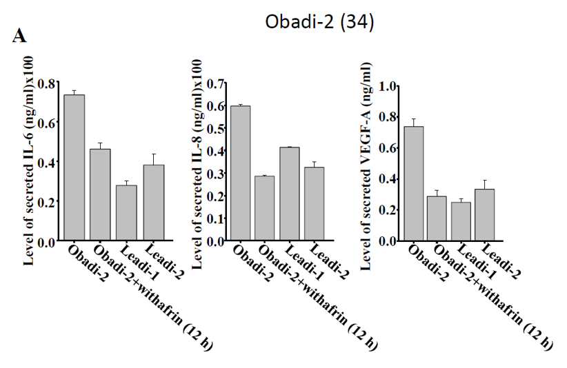
Furthermore, we assessed the levels of the secreted adipokines from Obadi and Leadi cells. To this end, serum-free conditioned media (SFCM) were collected from 2 Obadi cells treated as above and 2 Leadi cells, and then were utilized to assess the secreted levels of IL-6, IL-8 and VEGF-A by ELISA. Figure 2B and Supp. Fig. 1A show that Withaferin A significantly decreased the secreted level of these pro-inflammatory adipokines from Obadi cells to a level similar to that secreted from Leadi cells. Together, these data suggest that Withaferin A represses the expression/secretion of pro-carcinogenic/-inflammatory proteins in breast stromal adipocytes.
Active breast adipocytes from obese women are pro-angiogenic and Withaferin A represses this capability
Next, we sought to investigate the pro-angiogenic capability of Obadi cells and the effect of withaferin A, 2 Obadi cells were either mock-treated or treated with Withaferin A (250 nM), and were re-incubated for 24 hrs. 2 Leadi cells were utilized as control. Next, serum-free medium (SFM) was conditioned for 48 hrs with these cells. The resulting SFCM were added separately to 96-well plate seeded with HUVEC cells (1x10⁴) in matrigel and used for in vitro angiogenic assay. SFM was also added as negative control. Figure 3A and B show that after 5 hrs of incubation the number of HUVEC cells that were differentiated into closed cavities was significantly higher in the presence of SFCM from Obadi cells as compared to SFCM from Leadi cells. Interestingly, treatment with Withaferin A significantly decreased the number of closed cavities (Fig. 3A and B). This shows that Obadi cells are pro-angiogenic and Withaferin A can inhibit this activity.
To explore the molecular mechanism(s) that underlies the proangiogenic effect of Obadi cells, we assessed the level of proangiogenic proteins by immunoblotting. Figure 3C shows that the levels of vascular endothelial growth factor A (VEGF-A), hypoxia-inducible factor 1α (HIF-1α), and IL-8, were higher in Obadi cells as compared to leadi ones. Interestingly, treatment of Obadi cells with withaferin A decrease the levels of these proteins. These results show that the proangiogenic capability of breast stromal adipocytes is due to increase in the levels of the pro-angiogenic inflammatory proteins and that Withaferin A decreases their levels.
Next, the same SFCM collected above were used to examine the paracrine effect of Obadi cells on vascular formation in vivo. To this end, SFCM was mixed with matrigel and injected subcutaneously into nude mice. Eight days later, the matrigel plugs were extracted and imaged for the vascular formation. Figure 3D shows that while SFCM from Obadi cells promoted the vascular formation in vivo, as compared to Leadi cells, treatment of Obadi cells with withaferin A clearly suppressed the formation of blood vessels. Next, the level of CD31, as an endothelial cell marker, was assessed in the matrigel plugs by immunohistochemistry using anti- CD31 antibody. Figure 3E shows very low density of CD31-positive cells in matrigel plugs containing SFCM from Withaferin A-treated Obadi cells as compared to SFCM from control cells. In addition, we assessed the haemoglobin level, a surrogate for functional blood flow, in the matrigel plugs and showed that while SFCM from Obadi cells increased the concentration of haemoglobin as compared to SFCM from Leadi cells, SFCM from Withaferin A-treated Obadi cells significantly decreased the concentration of haemoglobin (Fig. 3F). This shows the pro-angiogenic effect of Obadi cells in vivo as well and withaferin A has the potential to reduce this capability.
Withaferin A represses Obadi-dependent stemness properties in luminal cells
We have previously shown that Obadi cells enhance the carcinogenesis of normal breast luminal cells through induction of the EMT process in a paracrine manner [4]. Therefore, we sought to investigate the effect of Withaferin A in repressing this pro-carcinogenic ability of Obadi cells on breast normal primary luminal cells. To this end, normal primary human breast luminal cells (NBL-10), were incubated for 24 h in serum-free conditioned medium (SFCM) collected from 2 Obadi cells treated either with DMSO or Withaferin A (250 nM) for 12 hrs. SFCM from Leadi cells was also utilized and SFM was used as negative control. The migration/invasion capabilities of these luminal treated cells were assessed using the xCelligence system. Figure 4A shows clear increase in the migration/invasion abilities of NBL-10 cells exposed to SFCM from Obadi cells as compared to SFCM from Leadi cells. Interestingly, SFCM from Withaferin A-treated Obadi cells significantly decreased the migration/invasion abilities of NBL-10, to a level similar to that observed in leadi cells.
Next, we investigated possible inhibition of mesenchymal features in luminal cells upon exposure to SFCM from Withaferin A-treated Obadi cells. Figure 4B shows that while the epithelial markers E-cadherin and EpCAM proteins decreased upon exposure of NBL-10 to SFCM from Obadi cells as compared to SFCM from Leadi cells, SFCM from Withaferin A-treated Obadi cells increased their levels similar to that observed upon exposure to Leadi cells. In contrast, while the mesenchymal markers N-cadherin was up-regulated in NBL-10 cells exposed to SFCM from Obadi cells as compared to SFCM from Leadi cells, SFCM from Withaferin A-treated Obadi cells decreased their levels similar to that observed upon exposure to SFCM from Leadi cells (Figure 4B). This indicates that while Obadi cells are pro-carcinogenic, treating these cells with Wthaferin A represses their activity.
Since EMT is a transition phase toward stemness, and the existence of links between the EMT and certain mammary stem cell properties, we investigated the possible effect of Withaferin A on Obadi cells-dependent stemness. Figure 4B and C show clear decrease in CD24 protein and mRNA upon exposure to SFCM from obadi cells as compared to SFCM from Leadi cells, while CD44 and ALDH1/2 were up-regulated as compared to their levels in luminal cells treated with leadi-SFCM or SFM. However, SFCM from withaferin A-treated Obadi cells significantly increased the level of CD24 and decreased the levels of CD44 and ALDH1/2 protein and mRNAs. This suggests the induction of stemness in breast luminal cells when exposed to SFCM from Obadi cells can be inhibited by treatment with Withaferin A.
Withaferin A represses the proangiogenic IL-8 by binding to its receptor CXCR1/2
To shed more light on the mechanism of Withaferin A-dependent repression of the procarcinogenic capability of Obadi cells, we investigated the ability of Withaferin A to inhibit the binding of IL-8 (as a pro-carcinogenic protein) to its receptors. To this end, we first assessed the levels of CXCR1 and CXCR2 proteins in whole cell extracts prepared from Obadi cells treated with Withaferin A for various periods of time. Figure 5A shows that the levels of both CXCR1 and CXCR2 significantly decreased upon treatment of Obadi cells with Withaferin A. Therefore, the decrease in the level of IL-8 protein upon treatment with Withaferin A is due to its binding to its receptors CXCR1/2. To test this hypothesis, the same protein lysates used above were used for immunoprecipitation using specific anti-CXCR1 antibody and specific anti-IL-8 antibody was utilized for immunoblotting. Anti-IgG antibody was used as a negative control. Figure 5B shows that concomitant to the decrease in the IL-8 and its receptor CXCR1 protein levels upon Withaferin A treatment, the level of IL-8-CXCR1 complex was also decreased in Withaferin A-treated cells as compared to their Sham-treated control cells. This indicates that Withaferin A is able to dissociate the biding of IL-8 to its receptors and hence represses the pro-carcinogenic capability of Obadi cells.
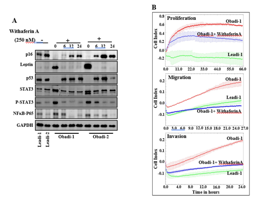
Figure 1: Withaferin-A normalizes the activity of Obadi cells. (A) Obadi-1 and Obadi-2 mature adipocytes were either sham-treated or challenged with Withaferin A (250nM) for the indicated periods of time. Cells were then collected and utilized to prepare the whole cellular lysates and used to assess the level of the indicated protein by immunoblotting using specific antibodies. GAPDH was used as internal control. Whole cell lystaes from Leadi-1 and Leadi-2 cells were utilized as control.
(B) Obadi cells were either sham-treated or challenged with Withaferin A (250nM) for 24 hrs and then were seeded either in complete medium in the E-plate to measure the proliferation rate for 72 hrs, or in SFM in the upper wells of the CIM plates, and the migration/invasion abilities were assessed for 24 h, by the RTCA-xCELLigence system.
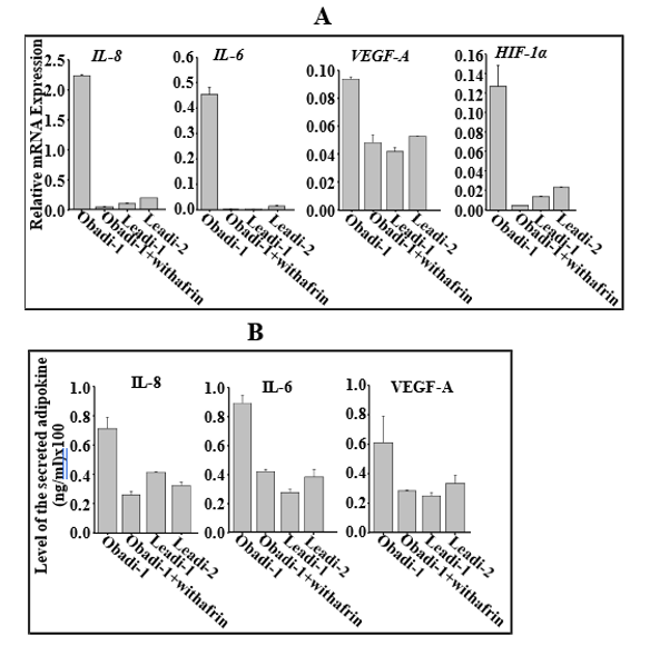
Figure 2: Withaferin A represses the expression/secretion of pro-carcinogenic/-inflammatory proteins in active breast stromal adipocytes. (A) Obadi-1 cells were either sham-treated or challenged with Withaferin A (250nM) for 24 hrs and then were collected and utilized to prepare total RNA to assess the level of the indicated transcripts by qRT-PCR. Total RNA extracted from Leadi-1 and Leadi-2 cells were utilized as control. Error bars represent means ± SD, values of 3 independent experiments. (B) SFCM from Obadi-1 cells treated as above was utilized to assess the level of the indicated adipokines by ELISA. SFCM from Leadi-1 and Leadi-2 cells were utilized as control. Error bars represent means ± SD values of 3 independent experiments. *P ≤ 0.00021.
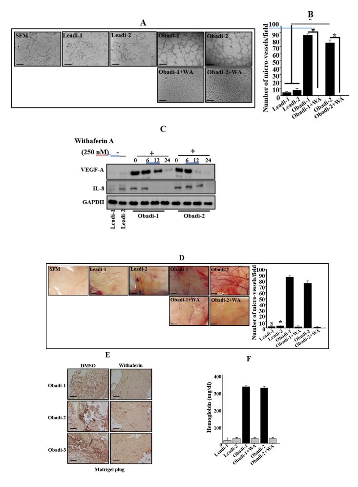
Figure 3: Active breast adipocytes from obese women are pro-angiogenic and Withaferin A represses this capability.
(A) SFCM were collected from the indicated cells and were applied independently on HUVEC cells plated on matrigel-coated 96-well plate, using SFM as negative control. The differentiation into capillary-like structures was assessed after 5 hrs of incubation. Representative photographs of HUVEC cavities are shown. Scale bars represent 30 μm. (B) Histogram shows average number of microvessels observed in five different fields. Error bars represent means ± SD. ***P
≤ 8.5x10⁻⁷. (C) Whole cell lysates were prepared from the indicated cells and were used for immunoblotting utilizing specific antibodies. (D) In-vivo matrigel plug assay. SFCM were collected from 48 hrs cultures of the indicated cells, centrifuged and mixed with phenol-red-free matrigel, and were injected subcutaneously into nude mice. Mice were then sacrificed after 8 days and the matrigel plugs were extracted for staining with the CD31 antibody (E), and for haemoglobin content (F).
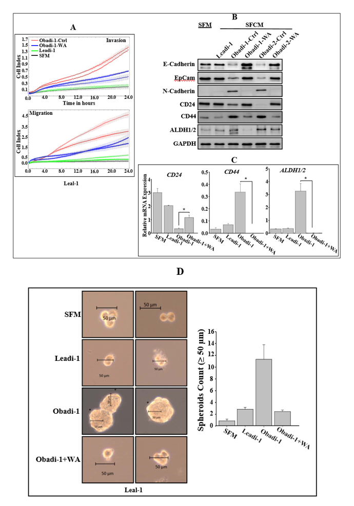
Figure 4: Withaferin A represses the pro-EMT and pro-stemness capabilities of Obadi cells.
(A) Obadi cells were either sham-treated or challenged with Withaferin A (250nM) for 24 hrs and then exposed to SFM for 24 hrs. SFCM were then collected. LeaL-10 luminal cells were exposed to either SFM as negative control, SFCM from Obadi-1 treated as above or SFCM from Lean-1 as control and then utilized to assess the migration/invasion and proliferation capabilities (the graphs are representative of different experiments), used to assess the levels of the indicated proteins by immunoblotting (B), or used to prepare total RNA to assess the level of the indicated transcripts by qRT-PCR (C). Error bars represent means ± SD, values of 3 different experiments. *P ≤ 2.33x10⁻⁵. (D). Cells (1000) were cultured in ultra-low attachment 96-well plates in the presence of specific stem cell medium. Left panel, representative images of mammospheres. Scale bar, 50 μm. Right panel, graphs depicting number of formed mammospheres. Experiments were performed in triplicate and several times; error bars represent means ± SD (*P < 0.05).
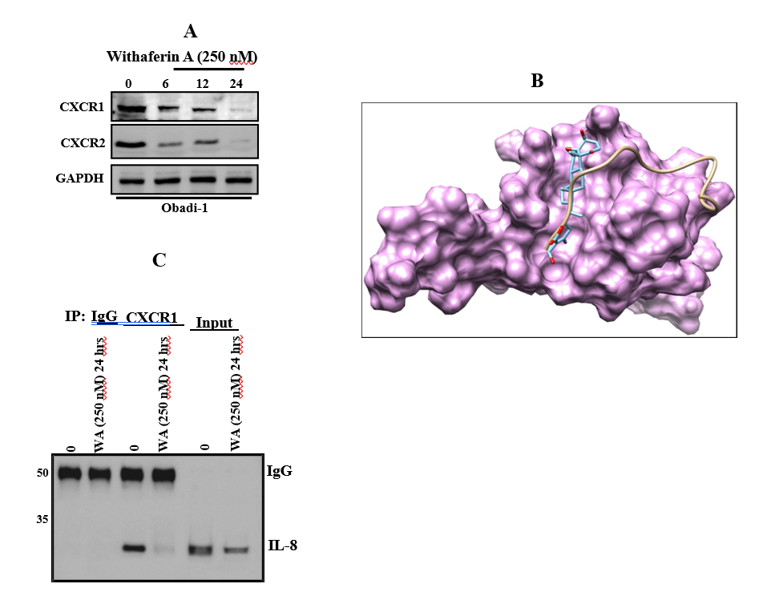
Figure 5: Withaferin A represses the proangiogenic IL-8 adipokine by binding to its receptor CXCR1/2. (A) Obadi-1 cells were either sham-treated or challenged with Withaferin A (250nM) for different periods of time and then were collected and utilized to prepare the whole cell lysates to assess the indicated proteins by immunoblotting utilizing specific antibodies. (B) The solvent-accessible surface of CXCR1 receptor (magenta) and stick rendering of bound IL8 (beige) and docked withafarin (cyan). (C) Obadi-1 cells were either sham-treated or challenged with Withaferin A (250nM) for 24 hrs and then were collected to prepare the whole cell lysates. 300 µg of the cellular extracts were utilized for immunoprecipitation using anti-CXCR1 antibody (mouse IgG was used as control). Immunoblotting was performed using specific IL-8 antibody. The input is the starting protein lysates was used as a positive control.
Discussion
Breast cancer progression is increasingly recognized as a multifactorial process driven not only by malignant epithelial cells but also by the surrounding tumor microenvironment (TME), particularly adipocyte-rich stromal compartments [7,8]. In addition, obesity is a serious risk factor for the development of breast cancer. We have shown recently that adipocytes from obese women are active and express low level of p16 [4]. Furthermore, we have demonstrated that p16 down-regulation activates breast stromal adipocytes and enhance their pro-carcinogenic effects in a leptin-dependent manner [4]. Additionally, we provide clear evidence that obesity induces EMT in breast epithelial cells in vitro and malignant transformation in vivo in a p16-dependent manner [4]. Therefore, we sought to find a strategy that can represses the activity of breast adipocytes derived from obese women. Our current findings provide mechanistic evidence supporting the role of adipocyte-derived inflammatory signaling in breast cancer progression and identify Withaferin A (WA) as a potential therapeutic modulator of adipocyte-derived inflammation.
In this study, we treated Obadi cells with minimal dose of WA and found that this treatment was able to restore the normal level of the p16 and leptin proteins similar to those found in Leadi cells (Figure 1). In addition, we demonstrated that active breast adipocytes significantly enhance angiogenic signaling through activation of the CXCR1/2–IL-8 axis, while treatment with WA effectively suppresses these pro-tumorigenic effects. The breast tissue microenvironment is highly enriched with adipocytes, which are now recognized as active endocrine and paracrine regulators rather than passive lipid storage cells [9]. Under pathological conditions, cancer-associated adipocytes (CAAs) secrete elevated levels of inflammatory cytokines, chemokines, angiogenic mediators, and extracellular matrix remodeling factors that collectively promote tumor growth, invasion, metastasis, and therapeutic resistance. Among these mediators, IL-8 has emerged as a critical inflammatory chemokine involved in angiogenesis, epithelial–mesenchymal transition (EMT), stemness, and metastatic dissemination [4,8]. The present findings reinforce previous observations demonstrating that IL-8 signaling contributes substantially to breast cancer aggressiveness, particularly within obesity-associated and inflammatory tumor microenvironments [8].
Our results further demonstrated that active breast adipocytes stimulate CXCR1 and CXCR2 signaling pathways, which are major receptors mediating IL-8 biological activity. Activation of the IL-8/CXCR1/2 axis has been associated with enhanced endothelial recruitment, neovascularization, tumor cell survival, and cancer stem cell maintenance [10]. Importantly, increased CXCR1/2 expression has been correlated with poor prognosis and increased metastatic potential in breast cancer patients [11]. The observed upregulation of angiogenic activity in our adipocyte-conditioned models suggests that adipocyte-derived IL-8 signaling may act as a central communication hub between stromal cells and tumor cells, facilitating a permissive microenvironment for tumor progression.
A major finding of this study is the potent inhibitory effect of WA on adipocyte-induced angiogenic and carcinogenic signaling. WA, a bioactive steroidal lactone derived from Withania somnifera, has gained increasing attention due to its anti-inflammatory, anti-angiogenic, and anti-cancer properties across multiple tumor models [12]. In our study, WA markedly suppressed IL-8 expression and attenuated downstream CXCR1/2 signaling, suggesting that its anti-tumor activity extends beyond direct cytotoxic effects on cancer cells to include modulation of stromal-tumor interactions within the TME. This is particularly important because targeting the supportive tumor stroma has become an emerging strategy in cancer therapeutics, especially in tumors characterized by chronic inflammation and obesity-associated signaling [13].
The anti-angiogenic effect observed following WA treatment may be mechanistically linked to the suppression of inflammatory transcriptional pathways such as NF-κB and STAT3, both of which are known upstream regulators of IL-8 expression [14]. Previous studies have demonstrated that WA interferes with NF-κB activation, leading to reduced cytokine production and inhibition of tumor-associated inflammation [15,16]. Therefore, it is plausible that WA disrupts the inflammatory feedback loop between adipocytes and tumor cells, thereby limiting endothelial activation and angiogenic remodeling. Moreover, suppression of CXCR1/2 signaling may also reduce cancer stemness and metastatic competency, given the established role of this pathway in maintaining breast cancer stem cell populations [17].
Another important implication of our findings relates to obesity-associated breast cancer progression. Obesity is characterized by chronic low-grade inflammation and adipose tissue dysfunction, resulting in increased secretion of IL-6, IL-8, TNF-α, leptin, and other pro-inflammatory mediators [18]. These alterations create a pro-tumorigenic microenvironment that accelerates disease progression and worsens clinical outcomes [19]. The ability of WA to suppress adipocyte-driven inflammatory signaling suggests potential therapeutic relevance in obesity-associated breast cancer, where conventional therapies often show reduced efficacy due to inflammatory resistance mechanisms [20].
The present study also highlights the importance of targeting stromal communication pathways rather than focusing solely on tumor cells. Increasing evidence suggests that therapeutic resistance and tumor recurrence are strongly influenced by reciprocal interactions between malignant cells and surrounding stromal components. By inhibiting adipocyte-mediated CXCR1/2/IL-8 signaling, WA may interfere with multiple hallmarks of cancer simultaneously, including angiogenesis, inflammation, invasion, and survival signaling. Such multi-targeted effects are particularly valuable in heterogeneous cancers such as breast carcinoma.
In conclusion, this study demonstrates that while activated breast adipocytes promote angiogenic and carcinogenic processes through activation of the CXCR1/2/IL-8 signaling axis, WA effectively suppresses these pro-tumorigenic effects. Further in vivo and translational studies are warranted to validate the therapeutic potential of WA in obesity-related breast cancer management. Our findings provide new insights into the mechanistic interplay between adipocytes and breast cancer cells and support the therapeutic potential of WA as a stromal-targeting anti-cancer agent. Targeting adipocyte-mediated inflammatory signaling may therefore represent a promising strategy for limiting breast cancer progression, particularly in obesity-associated disease settings.
Materials and Methods
Cells, cell lines, cell culture and chemicals
The adipose tissues were collected and immediately digested with collagenase to separate preadipocytes from the stroma vascular fraction, as previously described [4]. Preadipocytes were first cultured in special medium (PAGM) containing fetal calf serum (0.05 ml/ml), Endothelial Cell Growth Supplement (0.004 ml/ml), Epidermal Cell Growth Factor (10 ng/ml), Hydrocortisone (1 μg/ml) and Heparine (90 μg/ml), and used until confluency. Preadipocytes were then differentiated into mature adipocytes by replacement of the preadipocyte medium with serum-free preadipocytes differentiation medium (SFPADM), supplemented with D-Biotin (8 μg/ml), Insulin (0.5μg/ml), Dexamethasone (400 ng/ml), IBMX (44 μg/ml), L-Thyroxine (9 ng/ml) and Ciglitazone (3 μg/ml). Mature adipocytes were then cultured using adipocyte nutrition medium (ANM) containing fetal calf serum (0.03 ml/ml), D-Biotin (8 μg/ml), Insulin (0.5μg/ml), Dexamethasone (400 ng/ml). All supplements were purchased from PromoCell GmbH, Germany.
Normal breast luminal cells were obtained from the same tissues and were cultured as previously described [4].
Cellular lysate preparation and immunoblotting
This has been performed as previously described Antibodies directed against E-Cadherin (HECD-1), N-Cadherin, Epcam (VU1D9), NF-κB(p65), STAT3 and P-STAT3 were purchased from Cell Signaling Technology (Danvers, MA, USA). GAPDH (FL-335) from Santa Cruz (USA). p16 from BD Biosciences (San Jose, CA, USA). Leptin, IL-8, VEGF-A, CXCR1 and CXCR2 from Abcam (Cambridge, MA, USA). P53 (DO-1), ALDH1/2 (H-85) and CD24 (C-20) from Santa Cruz Biotech (Dallas, TX, USA), CD44 from Sigma-Aldrich (St. Louis, MO, USA).
RNA purification and quantitative RT-PCR
Total RNA, was purified using the mRNeasy mini kit (Qiagen, UK) according to the manufacturer's instructions and was treated with RNase-free DNase. 1 µg RNA was used to synthesize cDNA utilizing Advantage RT-PCR kit (Clontech Laboratories, Mountain View, CA, US). Quantitative RT-PCR was performed in triplicate using 4 µl cDNA mixed with 2x FastStart Essential DNA Green qPCR mastermix (Roche, New York, NY, US) and 0.3 µM forward and reverse primers. Amplifications were performed utilizing the LightCycler 96 Real-time PCR detection system (Roche) using the following cycle conditions: 95°C for 10 min (1 cycle); 95°C for 10 sec, 59°C for 20 sec, 72°C for 30 sec (45 cycles). GAPDH expression levels were used for normalization, and gene expression differences were calculated using the threshold cycle (Ct). The obtained values were plotted as mean ± SD. Three independent experiments were performed for each reaction. (See table 1 in the expanded view for the sequence of the primers).
|
Primers |
Sequences |
|
|
IL-8 |
Forward |
5’-GATCCACAAGTCCTTGTTCCA-3’ |
|
Reverse |
5’-GCTTCCACATGTCCTCACAA-3’ |
|
|
IL-6 |
Forward |
5’-AGACAG CCA CTC ACC TCT TCA G -3’ |
|
Reverse |
5’- TTC TGC CAG TGC CTC TTT GCT G -3’ |
|
|
VEGF-A |
Forward |
5’-CCCACTGAGGAGTCCAACAT-3’ |
|
Reverse |
5’-TTTCTTGCGCTTTCGTTTTT-3’ |
|
|
HIF-1α |
Forward |
5’-TCATCAGTTGCCACTTCCCCA-3’ |
|
Reverse |
5’-CCGTCATCTGTTAGCACCATCAC-3’ |
|
|
CD24 |
Forward |
5’-CAC GCA GAT TTA TTC CAG TGA AAC-3’ |
|
Reverse |
5’-GAC CAC GAA GAG ACT GGC TGT T-3’ |
|
|
CD44 |
Forward |
5’-CCA GAA GGA ACA GTG GTT TGG C-3’ |
|
Reverse |
5’-ACT GTC CTC TGG GCT TGG TGT T-3’ |
|
|
ALDH1A2 |
Forward |
5’-GAG TAA CTC TGG AAC TTG GAG GC-3’ |
|
Reverse |
5’-ATG GAC TCC TCC ACG AAG ATG C-3’ |
|
|
GAPDH |
Forward |
5’-GAGTCCACTGGCGTCTTC-3’ |
|
Reverse |
5’-GGGGTGCTAAGCAGTTGGT-3’ |
|
Table 1: List of the utilized qRT-PCR primers.
Immunohistochemistry
Formalin-Fixed/Paraffin-Embedded tissues (FFPE) were utilized for immunohistochemistry as previously described Tissues were incubated overnight with anti-CD34 antibody (Abcam), at a dilution of 1:500, followed by peroxidase-conjugated (Dako). Sites of antibody binding were visualized by the deposition of brown polymer of 3, 3′-diaminobenzidine chromogen (DAB) (Novocastra Laboratories Ltd, Buffalo Grove, IL).
Cell migration, invasion and proliferation
These assays were performed in a label-free real-time settings using the xCELLigence RTCA technology (Roche, Germany) that measures impedance changes in a meshwork of interdigitated gold microelectrodes located at the well bottom (E-plate) or at the bottom side of a micro-porous membrane (CIM plate 16) Cell migration and invasion were assessed as per the manufacturer’s instructions. In brief, 2x10⁴ cells in serum-free medium were added to the upper wells of the CIM-plate coated with a thin layer of Matrigel (BD Biosciences) basement membrane matrix diluted 1:20 in serum-free medium (invasion) or non-coated (migration). Complete medium was used as a chemo-attractant in the lower chambers. Subsequently, the plates were incubated in the RTCA for 24 h and the impedance value of each well was automatically monitored by the xCELLigence system and expressed as Cell Index (CI) value, which represents cell status based on the measured electrical impedance change divided by a background value. Each assay was performed in triplicate.
For the proliferation assay, exponentially growing cells (2x10⁴) were seeded in E-plate with complete medium as per the manufacturer’s instruction. Cell proliferation was assessed for 48h. All data were recorded and analyzed by the RTCA software. Cell Index was used to measure the change in the electrical impedance divided by background value, which represents cell status. Each assay was biologically performed in triplicate.
Immunoprecipitation
Cell were either sham-treated or exposed to Withaferin A (250nM) for 24 hrs. cells were then collected and utilized to prepare the whole cell lysates, which were then centrifuged at 14,000 rpm at 4°C. Subsequently, 300 µg of the cellular extracts were incubated with 2 μg CXCR1 antibody (Abcam) (mouse IgG1 was used as control) and mixed at 4°C for 4 h. Equal volume of protein A/G agarose was added per immunoprecipitation and mixed overnight at 4°C. After centrifugation, the pellet was washed 5 times with lysis buffer and placed in boiling water for 10 min in the presence of SDS gel sample buffer (0.5M Tris pH 6.8, 10% glycerol, 10% SDS, 5% 2mercaptoethanol, 1% bromophenol) and electrophoresed for 2 h at 125 V.
Conditioned Media
Cells were cultured in Serum-free media (SFM) for 48 h, and then the media were collected and centrifuged. Conditioned media (CM) were used either immediately or were frozen at -80°C until needed.
ELISA
ELISA was performed according to the manufacturer’s instructions. IL-6 and VEGF-A from R&D Systems (Minneapolis, MN, USA), IL-8 from Abcam (Cambridge, MA, USA). The OD was used at 450 nm on a standard ELISA plate reader (Bio-Rad, Hercules, California, USA). These experiments were performed in triplicate.
HUVEC endothelial tube formation assay
The formation of capillary-like structures was assessed in a 96-well plate coated with ice-cold growth factor-reduced Matrigel (in vitro angiogenesis assay, Millipore). After solidification of the matrix at 37ºC, 1x10⁴ HUVEC cells were seeded onto the polymerized matrix in the presence of 200 µl of conditioned medium. Formation of capillary-like structure was photographed after 5 hrs of incubation and their number was counted. The total tube area was obtained from five random microscopic fields and expressed as a mean of three different experiments.
In-vivo matrigel plug assay
Serum-free conditioned media (SFCM) were collected from 48 hrs cultures of Withaferin-treated Obadi cells and centrifuges to remove the cells. SFCM was then mixed with phenol-red-free matrigel (2:3 proportion, total 0.5 ml; BD Biosciences). Subcutaneous (s.c.) injection in mice was performed and the mice were killed after 8 days and the matrigel plugs were extracted for haemoglobin content (QuantiChromhaemoglobin assay; BioAssay Systems), and immunohistochemistry for CD34 antibody.
Statistical analysis
Statistical analysis was performed using GraphPad software. The results are presented as mean ±SD or SEM of at least three independent experiments. Statistical significance was determined using two-tailed Student’s t-test and p values of 0.05 and less were considered as statistically significant.
Funding
This work was supported by the Research Laboratory Department, King Faisal Specialist Hospital and Research Center under the RAC proposal # 2180022.
Disclosure statement
No potential conflict of interest was reported by the authors
Data availability statement
Supplementary data will be provided upon request.
References
- Kashyap D, Pal D, Sharma R, et al. Global Increase in Breast Cancer Incidence: Risk Factors and Preventive Measures. BioMed Res Int 2022, 9605439.
- Lee K, Kruper L, Dieli-Conwright CM, et al. The Impact of Obesity on Breast Cancer Diagnosis and Treatment. Curr Oncol Rep 21 (2019): 41.
- Zhang H, Wang M, Xu Y. Understanding the mechanisms underlying obesity in remodeling the breast tumor immune microenvironment: from the perspective of inflammation. Cancer Biol Med 20 (2023): 268-286.
- Al-Khalaf HH, Amir M, Al-Mohanna F, et al. Obesity and p16(INK4A) Downregulation Activate Breast Adipocytes and Promote Their Protumorigenicity. Mol Cell Biol 37 (2017).
- Atteeq M. Evaluating anticancer properties of Withaferin A-a potent phytochemical. Front Pharmacol 13 (2022): 975320.
- Lee J, Liu J, Feng X, et al. Withaferin A is a leptin sensitizer with strong antidiabetic properties in mice. Nat Med 22 (2016): 1023-1032.
- Wu C, Dong S, Huang R, et al. Cancer-Associated Adipocytes and Breast Cancer: Intertwining in the Tumor Microenvironment and Challenges for Cancer Therapy. Cancers 15 (2023).
- Al-Khalaf HH, Al-Harbi B, Al-Sayed A, et al. Interleukin-8 Activates Breast Cancer-Associated Adipocytes and Promotes Their Angiogenesis- and Tumorigenesis-Promoting Effects. Mol Cell Biol 39 (2019).
- Yu X, Zhang T, Cheng X, et al. Breast cancer cells and adipocytes in hypoxia: metabolism regulation. Discov Oncol 15 (2024): 11.
- Meier C, Brieger A. The role of IL-8 in cancer development and its impact on immunotherapy resistance. Eur J Cancer 218 (2025): 115267.
- Liu Q, Li A, Tian Y, et al. The CXCL8-CXCR1/2 pathways in cancer. Cytokine Growth Factor Rev 31 (2016): 61-71.
- Kumar S, Mathew SO, Aharwal RP, et al. Withaferin A: A Pleiotropic Anticancer Agent from the Indian Medicinal Plant Withania somnifera (L.) Dunal. Pharmaceuticals 16 (2023).
- Wang S, Hu H, Zeng W, et al. Targeting Tumor Stroma: Current Challenges and Future Directions. MedComm 7 (2026): e70763.
- Guo Q, Jin Y, Chen X, et al. NF-kappaB in biology and targeted therapy: new insights and translational implications. Signal Transduct Target Ther 9 (2024): 53.
- Heyninck K, Lahtela-Kakkonen M, Van der Veken P, et al. Withaferin A inhibits NF-kappaB activation by targeting cysteine 179 in IKKbeta. Biochem Pharmacol 91 (2014): 501-509.
- Rabiu L, Chinn M, Chen F, et al. Targeting Inflammation and Immune Regulation in Chronic Inflammation Associated Cancers. Cancer Sci 117 (2026): 1209-1222.
- Singh JK, Farnie G, Bundred NJ, et al. Targeting CXCR1/2 significantly reduces breast cancer stem cell activity and increases the efficacy of inhibiting HER2 via HER2-dependent and -independent mechanisms. Clin Cancer Res 19 (2013): 643-656.
- Gkrinia EMM, Belancic A. The Mechanisms of Chronic Inflammation in Obesity and Potential Therapeutic Strategies: A Narrative Review. Curr Issues Mol Biol 47 (2025).
- Yang Y, Ma X, Li Y, et al. The evolving tumor-associated adipose tissue microenvironment in breast cancer: from cancer initiation to metastatic outgrowth. Clin Transl Oncol 27 (2025): 2778-2788.
- Zhou Y, Bai M, Chen L, et al. Targeting inflammatory microenvironments: overcoming therapy resistance and immunosuppression. Mol Cancer 25 (2026).
Supplymentary Files